• Cataract Surgery: Couching to Phaco

    An exhibit celebrating the 50th anniversary of phacoemulsification and all the achievements in cataract surgery leading to it.
    Couching

    Couching is an ancient procedure for reclination of the cataractous lens. The earliest known description is found in the "Sushruta Samhita," published in 600 BCE in India. Knowledge of the surgery was likely spread via the Silk Road. Established in 130 BCE by the Han Dynasty in China, the Silk Road connected countries from Turkey to Japan for over a thousand years. Couching remained the most popular method of cataract surgery until the 19th Century.

    Georg Bartisch
    A black outlined woodcutting of a man performing an eye surgery. There are three men in the image who all wear Renaissance-style garments with ruff collars. The man performing the surgery holds a long needle in his hand, and he is inserting it into the left eye of the patient. The third man is wearing a hat, and he is holding the patients head still. Some boxes and bags can be seen hanging from a wall in the background.Georg Bartisch (1535-1606) was a surgeon who limited his practice to ophthalmology and hernia repair. In his book, "Ophthalmodouleia: das ist Augendienst" (1583) Bartisch describes how to fashion a cataract needle and perform couching. He warns patients away from itinerate surgeons at the market place. Instead he suggests surgery should take place inside a “light chamber in which the patient shall have his bed and covers; so that he need not go far. The nearer the bed, the better it is.”


    The Indian Operation
    A black and white photograph of a person performing an eye surgery similar to the image above. There are three people in the photo who all have dark skin and white hair. The person performing the surgery squats with their back to the camera, and is inserting a long needle into the eye of the patient, who is sitting on the ground. The third person is behind the patient, holding their head still.In "The Indian Operation of Couching for Cataracts" (1917) Robert Henry Elliot (1866-1936) describes documents several hundred cases of couching that he and other British surgeons observed in colonial India. Elliot calls the procedure “pagan” and states that Westerners “oft-expressed wonder...not that their results are so bad, but that they are ever good.”
    Extracapsular Cataract Extraction (ECCE)

    Jacques Daviel (1696-1762) performed the first planned extracapsular cataract extraction on a living human eye in 1750. Three years later, Samuel Sharp reported on intracapsular cataract extraction. The debate about whether to perform extractions led to heated exchanges between ophthalmologists, leading Michel de Wenzel in 1791 to lament, “Why do [surgeons] not agree among themselves, which mode is most simple and easy; and which the most likely to procure success?”

    Daviel's Operation

    A pencil drawing of four different medical instruments. The top drawing shows a hand holding a knife with a small, flat blade and a long handle. The knife is being inserted into a drawing of a human eye. The other drawings are of two similar knives without a hand holding them and one pair of black medical scissors.Extracapsular cataract extraction, as described by Jacques Daviel, required several instruments including keratome, knife, spatula, scissors, needle and spoon. According to Julius Hirschberg’s "History of Ophthalmology," “The incision was made near the lower limbus, because of the patient’s tendency to roll the eyes upward.”
    Intracapsular Cataract Extraction (ICCE)

    Intracapsular cataract extraction (ICCE) was introduced in 1753 by Samuel Sharp. It was generally dismissed until the 20th Century because it was considered incredibly difficult when compared to the relatively simple procedure of Daviel. Lt. Col. Henry Smith (1859-1948) of India is credited for making ICCE popular again at the turn of the last century.

    Capsule Forceps

    A pair of silver-colored metal tweezers. They have long, needle-nose ends and they sit on a blue background.Capsule forceps like this example was used to grasp the lens capsule and gently pull it from the eye with a side to side motion that broke the zonular insertion.


    Phacoerysis

    A silver-colored metal medical tool sits in a hinged case lined in bright red cloth. The tool is made of three small cylindrical parts, and the gold text on the case lid reads: Jose Vilaplana, BarcelonaIgnacio Barraquer, MD (1884-1965) introduced suction to cataract surgery in 1917, calling the procedure phacoerysis. Arguing for his technique Barraquer once said, “compare the [use of] forceps to the claw of a cat and the cup to the lips of a beautiful woman. What would you prefer to feel on your cheek?”


    Cryo-Extraction

    An illustrated advertisement for a medical tool. The background of the ad is brown and features a drawing of doctors in surgery. The foreground shows a hand holding a white plastic tool and inserting it into a drawing of a human eye. There is small white text on the ad that is too small to read, but the white text in the center reads: F-20/20 Cryoextractor.Tadeusz Krwawicz, MD (1910-1990) introduced cryo-extraction of the cataractous lens in 1961. Many designs for cryo-extractors quickly followed suit including this instrument.


    Sutures

    The first known use of sutures in cataract surgery was by Henry Willard Williams (1821-1895) in 1865. Their use was more universally accepted, however, after the introduction of topical anesthesia in 1884. In 1891 Ferdinand Suarez de Mendoza described the first preplaced sutures.

    For Sutures

    A white and black paper box with two large curved needles laying next to it. The box has writing in French on it, including a title which reads: Aiguilles de Chirurgie. The needles are long and curved, but very thin.Physicians devised a variety of sutures which were believed to promote healing and provide, as Hasket Derby put it, protection from “excessive coughing, vomiting, sneezing, etc. and even from such accidents as falling out of bed.”


    Against Sutures

    An black and white illustration of a human eye being held open by four metal wires. There are small black lines around the iris of the eye mimicking stitches or sutures.In the late 20th Century, physicians began to question the use of sutures, such as this comment by Leon Alger in 1971: “Sutures in cataract surgery are of such variety as to make one wonder if any given suture is entirely satisfactory…One can imagine the patient’s discomfort for weeks and months while he winks and blinks and nods until these miserable things are finally gone.”


    Anesthesia

    Ophthalmologists were aware of general anesthesia but rarely used it because of side effects such as nausea and retching, as well as their desire to keep patients responsive during surgery. In 1882 Hasket Derby wrote “anesthesia is…only necessary in the case of very nervous patients or for those who lose their self-control when the speculum is applied.”

    Carl Koller
    A sepia-toned photograph of a young man with a mustache. He is a white man with curly dark hair parted in the middle, and he has a long pointed mustache. He wears a dark suit and looks directly into the camera.Carl Koller (1857-1944) announced topical anesthesia in 1884, revolutionizing cataract sugery. Many physicians immediately recognized its importance. Herman Knapp, for instance, hailed it as the “greatest gift we have received for many years.”


    Hasket Derby
    A silver-colored mesh mask made of metal. The mask has a handle and an oval mouthpiece , and there is wire mesh over the mouthpiece.Hasket Derby wrote an entire book about general anesthesia and cataract surgery in 1882 in which he noted: “the American patient has been in the habit of demanding artificial insensibility to pain to an extent unusual in other countries, and indeed, unjustifiable…”
    Intraocular Lenses (IOLs)

    After WWII, Dr. Harold Ridley (1906-2001) was operating on a cataract patient when a resident remarked how it was a pity not to be able to replace the removed lens. This comment was the impetus for Ridley’s innovation. He later remarked that cataract extraction was “but halfway to a cure.”

    The First Implant
    A small, round plastic medical device sits on a red foam lining inside a white, round plastic case.The first intraocular lens implant surgery was performed on November 29, 1949. Ridley used extracapsular cataract extraction because the original IOL was designed to be placed in the posterior chamber.


    IOL Design

    A black and white illustration of six small, round medical devices. They each have a circular center, and they each have two handles or flaps, but the shapes of those handles differ on each of the six drawings.Ridley’s original design for the intraocular lens was not optimal. Many tried to find the perfect solution by creating posterior chamber, anterior chamber and iris fixation lenses. The IOL finally received FDA approval in 1980.
    Phacoemulsification

    Charles Kelman, MD (1930-2004) famously had an “aha” moment when a dentist used a Cavitron high-frequency ultrasonic probe to clean his teeth. The dental probe had to be significantly modified, but the phacoemulsification procedure was ready for the first human patient in 1967. Two of his colleagues talk about his influence on cataract surgery in this video.

    Mastering Phaco
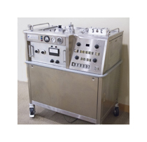
    A large, silver-colored metal machine on wheels. The machine has two handles on the top and two wheels on the bottom, and has many black buttons and switches.In the early years many surgeons found phacoemulsification difficult to master. Kelman recognized that surgical outcomes depended heavily on proper use of the machinery so he provided instruction courses starting in the early 1970s.


    Phaco and IOLs
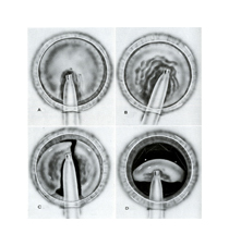
    Four black and white illustrations of a medical tool entering a circular structure. The syringe-shaped tool appears to be sucking out the fluid on the inside of the circle, and each image shows more of the fluid disappearing.There was limited incentive for small incision surgery when the wound had to be opened from 3.0 mm to 7.0 mm in order to accommodate the early intraocular lens implants. It would take over 10 years before foldable IOLS and familiarity with microsurgery made phacoemulsification the procedure of choice.