• Military Ophthalmology

    The Truhlsen-Marmor Museum of the Eye presents an exhibit exploring the relationship between ophthalmology and the armed forces - uncovering the stories that intersect medicine and the military.

    Military Medicine

    Military medicine keeps armed forces healthy during peace and heals soldiers after conflict. While we often associate military medicine with battlefield surgery, it also encompasses sanitation, transportation, and rehabilitation. 

    Napoleonic Wars (1798-1815) and Ophthalmology 

    The Napoleonic Wars exposed soldiers from around Europe to an eye disease known as “military ophthalmia” which was, in fact, several different types of bacterial infections. These diseases were brought home by returning soldiers where they were studied by physicians such as John Vetch who published a seminal study on the causes of trachoma in 1807 (pictured).


    Military Hospitals

    A black and white photograph of a group of soldiers. One soldier is performing an exam for eyeglasses on another solider. The man performing the exam is wearing a uniform and black leather boots, and he is pulling lenses from a large case on a table. The man receiving the exam is wearing a large military overcoat and has a pair of glasses with empty frames to hold test lenses. Seven other men in military uniforms sit behind them.This image is from a pamphlet distributed in 1920 describing U.S. Base Hospital No. 115 established in Vichy, France during WWI. Pictured is part of their eye clinic.


    Army Medical Museum

    The Army Medical Museum was founded in 1862 during the American Civil War. It was created to recover medical specimens from the battlefield, catalogue, and analyze them to improve the effectiveness of military medicine. The collection grew in subsequent years with an emphasis on pathological specimens and medical photography. In 1918, Dr. Harry Gradle (pictured) agreed to send eye pathological specimens to the Army Medical Museum in exchange for help in their analysis. The resulting Registry of Ophthalmic Pathology became the first of several specialty divisions created at the museum. Eventually Helenor Campbell Wilder was hired to develop and manage the ophthalmic registry and, in 1949, the Army Medical Museum was renamed the Armed Forces Institute of Pathology (AFIP). For years the Army Medical Museum collaborated openly with non-military pathologists who reviewed and diagnosed cases. The partnership lasted because of the mutual interest of military and civilian medicine in advancing technical processes and interpreting pathological specimens. The AFIP closed, and the specimens moved to the National Museum of Health and Medicine, in 2011.

    For more about Ms. Wilder and women in ophthalmology, visit the Remarkable Women exhibit.
    Test for Color Vision

    Shinobu Ishihara attended the Imperial University in Tokyo under a full scholarship from the Japanese army. After earning his medical degree, he trained for two years with Professor Komoto in ophthalmology and then began teaching at the Military Medical School. The army sent Ishihara for further ophthalmic training with Professor Wolfgang Stock in Germany; he returned to Japan in 1914 at the outbreak of World War I. At that time, he was assigned the task of creating a color vision test for army recruits. The first Ishihara plates were issued by the Japanese army in 1916. 

    Shinobu Ishihara, MD (1879-1963)
    Dr. Ishihara personally edited each edition of his book because the color variations had to be exact. After his death, his daughter Mineko carried on this tradition until her death in 1969. 


    Tests for Color Blindness
    An example of the Ishihara color blindness test that uses Arabic numerals.Dr. Ishihara created his test so that everyone could see something on the page - making it impossible to cheat or intentionally fail. The original army tests in 1916 used Japanese hiragana characters (more cursive). In the same year, Ishihara published a set of plates for wider distribution, using Japanese katakana characters (more angular). Finally, an international edition was published using Arabic numerals in 1919 and distributed overseas.
    Positive Black Out

    In 1951, Thomas Duane was stationed at the Naval Air Development Center - home of the Johnsville human centrifuge. The centrifuge had been installed the year before and was the world’s most powerful tool for studying the effects of g-forces on the human body. Pilots can black out while experiencing positive g-forces during flight. Duane’s study determined why this happened and documented “greyout,” when vision is dimmed, and peripheral vision lost before a pilot blacks-out or faints due to loss of blood pressure. Emil Bethke was then brought in to illustrate Duane’s findings. It would be 15 years before a fundus camera was mounted to the centrifuge to photograph the phenomenon.

    Thomas Duane, MD (1917-1993)
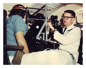
    The centrifuge remained in use for almost 50 years as part of training for pilots and astronauts. John Glenn recalled, “[In the centrifuge] you were straining every muscle of your body to the maximum…if you even thought of easing up, your vision would narrow like a set of blinders, and you’d start to black out.”


    Bethke's Retinal Illustrations

    According to Duane, medical illustrator Emil Bethke spent only “about a week” with the study “but during that time rode the centrifuge time and time and time again, which was a huge undertaking in itself, until he finally was able to see the changes going on in the subject’s eyes.” Afterwards, Bethke produced nine illustrations for Duane.


    The Intraocular Lens Implant

    Sir Harold Ridley first came into contact with polymethyl methacrylate (PMMA or Perspex) while treating fighter pilots during World War II. The plastic was used in the windshield and canopies of two airplanes being flown by the Royal Air Force. Ridley observed that PMMA was intert and well-tolerated by the body, but his ideas for the material coalesced only after the war when a resident remarked that it was a pity not to be able to replace the removed lens. After meeting with Rayner, Ltd., Ridley introduced the world's first intraocular lens, which he surgically implanted on November 29, 1949. In honor of his services to cataract surgery, Dr. Ridley was conferred a knighthood by Queen Elizabeth II in February 2000. 

    Sir Nicholas Harold Lloyd Ridley (1906-2001)

    Dr. Ridley once remarked, "Immediately after a major war many were accustomed to accepting risk of injury. If implants had not started in 1949, they might have been delayed for yet another forty years."


    IOL Implantation

    These film stills document the eighth IOL surgery performed by Ridley and the second to be filmed.