OCT 31, 2016
By John Lillvis, MD, PhD, and Elias I. Traboulsi, MD
A Compendium of Inherited Disorders and the Eye, Oxford University Press
Genetics
OMIM Numbers
Inheritance
Gene/Gene Map
- 1-2 Mb deletions of chromosome 7q11.23 are observed in 95% of individuals with clinical features of Williams syndrome. Approximately 28 genes have been mapped to this region, including elastin (ELN), LIM kinase-1 (LIMK1), replication factor c, subunit 2 (RFC2), GTF2I repeat domain-containing protein 1 (GTF2IRD1), and general transcription factor III (GTF2I). Hemizygosity of the ELN gene has been implicated in the cardiovascular phenotype, and hemizygosity of GTF2IRD1/GFT2I has been suggested as the basis for the neurocognitive features of the disorder.
Epidemiology
- Prevalence is 1 in 7,500-10,000 children.
Clinical Findings
There are three recognized clinical occurrences of aniridia:
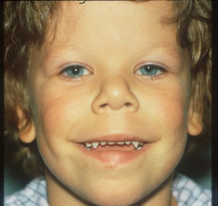
- The full-blown form of the syndrome includes supravalvular aortic stenosis, multiple peripheral pulmonary arterial stenoses, elfin face, mental and statural deficiency, characteristic dental malformation, and infantile hypercalcemia.
- Significant phenotypic heterogeneity with respect to the presence and severity of clinical features is characteristic.
Ocular Findings
- Holmstrom et al examined external eye photographs of 43 children with the syndrome and 124 control subjects. A stellate pattern was noted in the irides of 51% of the Williams patients and 12% of controls. The pattern was easier to detect in lightly pigmented irides.
- In a study of 152 patients, Winter et al found that 54% had strabismus and almost all had esotropia. In Europeans, blue irides were present in 77%, green in 7%, and brown in 16%. A typical stellate iris pattern of the anterior stroma was found in three-fourths of patients. Wolfflin-Krichmann whitish spots were also detected in brown irides. Retinal vascular tortuosity was found in 22% of patients, and 3 patients had cataracts. There were no ocular abnormalities that could be attributed to hypercalcemia.
- A smaller case series of 16 patients by Viana et al found that slightly higher rates of stellate irides (81.2%) and retinal vessel tortuosity (37.5%), but a lower rate of strabismus (18.7%). Patients with cataract (1/16), megalocornea (1/16), keratoconus (1/16) and optic disc hypoplasia (2/16) were also reported.
- Case reports of abnormal posterior insertion of the extraocular muscles (Ali SM and Shun-Shin GA; and Bela C and Klainguti G), keratoconus (Pinsard et al), anterior segment dysgenesis (Todorova et al) and rod-cone dystrophy (Kuehlwein L and Sadda SR) in patients with Williams syndrome of have been reported.
Therapeutic Aspects
- Esotropia and amblyopia are treated as appropriate with surgery and patching.

Figure 1. Typical facial appearance of a patient with Williams syndrome
References
- Ali SM, Shun-Shin GA: Abnormal extraocular muscle anatomy in a case of Williams-Beuren Syndrome. J AAPOS. 2009; 13:196-197.
- Bela C, Klainguti G: Abnormal extraocular muscle insertion in Williams Beuren syndrome (WBS). Klin Monbl Augenheilkd. 2014; 382-383.
- Holmström G, Almond G, Temple K, Taylor D, Baraitser M: The iris in Williams syndrome. Arch Dis Child. 1990; 65:987-989.
- Kuehlewein L, Sadda SR: Rod-cone dystrophy associated with Williams syndrome. Retina Cases Brief Rep. 2015 Fall; 9(4):298-301.
- Pinsard L, Touboul D, Vu Y, Lacombe D, Leger F, Colin J: Keratoconus associated with Williams-Beuren syndrome: first case reports. Ophthalmic Genet. 2010; 31:252-256.
- Strømme P, Bjørnstad PG, Ramstad K: Prevalence estimation of Williams syndrome. J Child Neurol. 2002; 17:269-271.
- Todorova MG, Grieshaber MC, Cámara RJ, Miny P, Palmowski-Wolfe AM: Anterior segment dysgenesis associated with Williams-Beuren syndrome: a case report and review of the literature. BMC Ophthalmol. 2014; 14:70.
- Viana MM, Frasson M, Galvão H, et al: Ocular Features in 16 Brazilian Patients with Williams-Beuren Syndrome. Ophthalmic Genet. 2015; 36:234-238.
- Winter M, Pankau R, Amm M, et al: The spectrum of ocular features in the Williams-Beuren syndrome. Clin Genet. 1996; 49:28-31.
Resources
- Williams Syndrome Association 570 Kirts Boulevard, Ste. 223 Troy, MI 48084-4156 248-244-2229 | 800.806.1871 info@williams-syndrome.org. www.williams-syndrome.org.
- The Lili Claire Foundation 3540 West Sahara Ave., #182 Las Vegas, NV 89102 (702) 862-8141 Staff@LiliClaire.org http://liliclaire.org
Traboulsi EI. Compendium of Inherited Disorders and the Eye. New York: Oxford University Press; 2005. Adapted with permission.