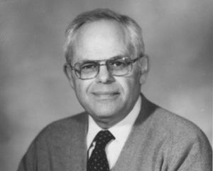
I first became aware of Ben Fine, MD, when as a first-year resident in pathology, I heard him speak in December 1962 at an Association for Research in Vision and Ophthalmology (ARVO) meeting, then held in Ann Arbor, Mich.
Dr. Fine presented a paper on the electron microscopic anatomy of the eye. Two things struck me. First, he was one of the most articulate speakers that I had ever heard. Second, he took a profoundly complex, potentially boring subject and told a captivating, precise story that not only unraveled the complexities, but did so in a way that made it imminently understandable and totally interesting.
 I next saw and met Dr. Fine in July 1964 when I started my fellowship at the Armed Forces Institute of Pathology (AFIP) in Washington, D.C. Dr. Fine was a Canadian citizen, from Ontario. He received his undergraduate and medical training at the University of Toronto, Toronto, Ontario, graduating in 1953. From 1953 to 1956, he was an intern and then a resident in ophthalmology at the District of Columbia General Hospital, Washington, DC, followed by a fellowship in ophthalmic pathology with Lorenz E. Zimmerman, MD, at the AFIP.
In 1964, Dr. Fine spent half time on the faculty at the AFIP and the other half time as a practicing, clinical ophthalmologist, positions that he maintained until he retired. His office and laboratory were across the hall from where the AFIP fellows and the group’s director of the ophthalmic pathology division, Dr. Lorenz Zimmerman, were housed.
Dr. Fine’s doors always were open and welcoming. He had the perspective both of a scientist and of a clinician, attributes rarely found in one person. One of the many remarkable things that he possessed was his ability to interact with the fellows as a mentor, having total patience to listen and then to give meaningful, thoughtful, practical advice. I had already had two years of a pathology residency and was set to start my residency in ophthalmology in July 1965. We spent long hours talking about how to have a career combining ocular pathology and clinical ophthalmology. These were hours well spent. Dr. Fine was my mentor but also became my lifelong friend.
In 1964-65, ocular electron microscopy was in its infancy. Few scientists were involved in electron microscopic interpretation of ocular tissues, both normal and pathologic; fewer still had the expertise to interpret fully what they were finding. Dr. Fine truly was a pioneer and the premier electron microscopist at that time and was the person mainly responsible for laying the groundwork and principles as we know them now. Not only did he do this for the normal anatomy of ocular tissue, but he performed this service for pathologic conditions as well, e.g., corneal pathology.
Dr. Fine had many remarkable attributes, but probably the most obvious one was his ability to listen and then advise, always in a calm, unflappable, even tone. I never saw him angry. He had a calmness about him that was remarkable. There was only one time did I see that calmness develop some cracks. My wife had family from Canada, some of whom were hard of hearing.
One evening during my year at the AFIP, we went to a cocktail party where my wife met Dr. Fine for the first time. As she was talking to him, Dr. Fine turned to hear better, explaining that he had loss of hearing in his left ear. My wife, thinking of her Canadian family with loss of hearing, jokingly said to Dr. Fine, “Oh, you must be from Canada.” Dr. Fine, startled, stated, “How did you know?” My wife, now equally startled, explained about her Canadian family. It was one of the few times that I ever saw Dr. Fine lose his calm.
During the year that I was at the AFIP, we agreed that we would write two books, first, Ocular Histology (that went through 2 editions) and second, Ocular Pathology (that went through 5 editions). In those days no computers or digital photography existed. Writing a book was a long, difficult process. Text was typewritten. All illustrations were prepared, cropped, and put together manually. Although this was planned in 1965, Ocular Histology was not published until 1972 and “Ocular Pathology: A Text and Atlas” in 1975. What was remarkable was that in the preparation of the seven editions, Dr. Fine and I never had an argument over content, meaning, illustrations and so forth. If we had a disagreement, a rare occurrence, we simple figured it out in a calm, logical way.
Dr. Fine wrote over 100 articles in peer-reviewed journals. The articles were diverse and often groundbreaking. Some examples of the papers are:
Glutaraldehyde fixation of pathologic and surgical ocular tissues (showing that glutaraldehyde could be used as a primary fixative, especially if electron microscopy was scheduled)
Diabetic lacy vacuolization of iris pigment epithelium (first example and showed clinical correlation)
The ocular histology of homocystinuria
A light and electron microscopic study (shown for the first time)
Marfan syndrome. A histopathologic study of ocular findings (shown for the first time)
Hemoglobin SC retinopathy and fat emboli to the eye
A light and electron microscopical study (shown for the first time)
Lattice corneal dystrophy
Report of an unusual case (actually this was the first case of the Avelino type, which had not yet been described)
Meesmann's epithelial dystrophy of the cornea (beautifully illustrated)
A clinicopathologic study of four cases of primary open-angle glaucoma compared to normal eyes (proposed a very plausible cause of the glaucoma)
Histopathology of transient neo-natal lens vacuoles (shown for the first time)
He loved his nonsurgical clinical practice. He said that it kept him balanced. Conversely, he thought that his laboratory work, his teaching, and his mentoring kept him sane. But most of all, he loved his family. He was a devoted husband to his wife Frumie and father to his daughters Nina and Sharon. Here are their observations:
Sharon: “He was the only father who let his 10-year-old daughter use a full-scale electron microscope. I remember visiting the AFIP where he would teach me about all the equipment (I secretly would have preferred a pony). He had a lot of interesting summer jobs growing up, including as a packager at a salmon canning factory, as well as an ice cream packager. Nina recalls him telling her that the workers were allowed to eat the broken ice cream bars, and so sometimes he would accidentally on purpose drop a crate. When people complemented him on his brilliance or used the word ‘genius’, he would smirk and say, ‘Genius is 99% perspiration and 1% inspiration, so you will always have to do the work.’ Or you might hear him say, ‘The eyes are the brain’s way of coming out to play.’ He was a playful father and had a great sense of humor.”
Nina: “He was a very strong, smart, funny, charming man and a sweet caring father. Always telling funny jokes or pretending to sing badly. He loved word play, and this would put a big smile on his face. It was very important to him to provide a good life for his family, and he worked very hard to do so, and he succeeded. His own family in Canada struggled through the Great Depression, and so he always was frugal with money, making sure we always had enough, including funding for both kids to go to college and even graduate school. He was kind to our mother's family, always making Friday night dinners at the farm a priority and even developing a special bond with our mother's brother, Kopel, after her death. Most people would have described [Dad] as a teddy bear, and I can remember his warm hugs.”
Dr. Fine was my role model. I loved him as an older brother. His crystal-clear reasoning still reverberates in my head. The later years were not kind to him. He had heart disease with quadruple bypass surgeries at age 50, and then redoing them all at age 51 or 52, probable strokes along the way, some angioplasties and finally an emergency bypass again at 68, where he was on a respirator in a semi-coma for a long while. It sent him into a decline and into a state that eventually mimicked Parkinson’s disease.
He was a giant in our field, laying down the foundations of electron microscopy for future generations. Dr. Fine is still missed, and we will never forget him.
Editor’s Note: We are grateful to our History of Ophthalmology editor, Daniel M. Albert, MD, MS, and his editorial assistant, Ms. Jane Shull, who contributed to the editing of this article.