Cataract formation is a common complication in eyes with uveitis. This is typically due to chronic intraocular inflammation, long-term corticosteroid use or both. In addition, cataract formation in uveitic eyes tends to occur at a younger age than senile cataracts. It may also occur with other complications, such as posterior synechiae, anterior synechiae, band keratopathy, pupillary membranes and fibrosis of the anterior capsule. Therefore, cataract surgery in uveitic eyes can be associated with significant intraoperative and postoperative complications, suggesting high risk and poor prognosis. However, these 10 pearls will help you achieve successful anatomic and visual outcomes.
1. Stay calm and expect the unexpected. In addition to your typical cataract tray and phacoemulsification machine, you should plan ahead for difficulty at any and every step of the surgery. Alert your operating room staff to the things you want to have in the room just in case. I recommend trypan blue, iris hooks, retinal scissors, capsular tension rings, anterior vitrectomy and a lens loop.
2. Wait for a period of documented quiescence before surgery.Achieve remission by any means possible, whether that is local or systemic treatment. I recommend at least three months of quiescence, but in severe cases, a longer duration without flare-ups is preferable. During this period, regularly monitor the patient to confirm the lack of an anterior chamber cell, retinitis, vitritis and cystoid macular edema. Exceptions to this rule are phacoantigenic uveitis and pediatric cases at immediate risk for amblyopia. Any infectious cause of uveitis should be completely addressed prior to surgery.
3. Give perioperative steroids. Do not make any drastic changes to your patient’s treatment regimen immediately prior to surgery. If the patient has a rheumatologic disorder that is well controlled on a stable dose of immunosuppression, do not discontinue the drug prior to surgery. Consider treating patients with severe systemic or ocular autoimmune disease with a short course of high-dose oral corticosteroids a few days prior to surgery and into the immediate postoperative period. Any patient taking chronic systemic corticosteroids should also receive a stress dose of corticosteroids on the day of surgery.
4. Don’t use topical anesthesia alone. Intraoperative complications can occur in uveitic eyes, even in seemingly straightforward cases. Iris manipulation is uncomfortable for the patient. The use of a peribulbar or retrobulbar block is sufficient in most cases. However, general anesthesia is necessary for young pediatric patients and prolonged cases, such as those combined with glaucoma or retinal surgery.

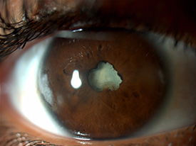
Posterior synechiae and a dense cataract in a HLA B27 positive patient. Photo courtesy of Dr. Lee.
5. Use a viscoelastic cannula to perform synechiolysis.Introduce the viscoelastic cannula under a nonadherent area of the pupillary border, preferably through a paracentesis wound approximately 180 degrees away from the posterior synechiae. Sweep laterally to lyse the adhesion at the pupillary margin. This simple maneuver will easily achieve synechiolysis if the lens capsule is only adherent to the pupillary margin. Then, you can use viscoelastic to perform viscomydriasis. In more severe cases of posterior synechiae, perform a careful preoperative slit-lamp exam to find any tiny openings in the pupil from which to position the cannula. Beware of adhesions between the posterior iris stroma and anterior capsule extending beyond the pupillary margin. With extensive iridocapsular adhesions, be careful not to tear the iris or puncture the lens capsule; a Kuglen hook is helpful to perform gentle dissection in these cases.

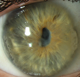
Pupillary membrane in a patient with uveitis associated with juvenile idiopathic arthritis. Photo courtesy of Dr. Lee.
6. Beware of a pupillary membrane occluding the pupil. Usually, the pupillary membrane is easily removed at the time of posterior synechiolysis. However, in cases of prolonged intraocular inflammation, this membrane can be thick and fibrotic, making it difficult or impossible to disrupt without a sharp instrument. Take caution due to the possibility of bleeding and iridodialysis with forceful manipulation. Once an opening in the membrane is made, retinal scissors can be used to cut the membrane without any tension on the iris.
7. Be prepared for a floppy iris. Pupillary dilation is difficult in uveitic eyes due to the presence of posterior synechiae and atrophy of the iris sphincter. There is usually little or no response to pharmacologic dilation. After posterior synechiolysis and pupillary membrane excision, you can attempt viscomydriasis. Do not be surprised if the iris begins to behave like the worst Flomax-related intraoperative floppy iris syndrome you’ve ever seen, even with the use of intracameral epinephrine. Iris hooks, either a single hook or several, are your best friends in this situation. A 27-gauge needle is an efficient way to create small paracentesis wounds for hooks (these do not require hydration at the end of the case). It is easier to engage all the hooks with gentle tension before maximally retracting them individually.
8. Beware of a fibrotic anterior capsule. Due to the density of the lens and fibrosis of the anterior capsule, trypan blue is very helpful. Pupillary membranes can occur in multiple layers and can be confused with the anterior capsule. If you think you have completed your capsulorrhexis but cannot insert the hydrodissection cannula, consider restaining the capsule. You must have many capsulorrhexis techniques in your armamentarium, including use of the vitrector or retinal scissors to open the lens capsule.
9. Place a three-piece monofocal IOL or leave the patient aphakic.With advanced phaco technology and modern lenses, nowadays most uveitis patients receive IOLs. In young children or cases where inflammation is expected to be chronic and relentless, aphakia is an appropriate choice. Choose an IOL wisely. Multifocal and accommodative lenses are a poor choice in chronic uveitis because future episodes of inflammation may compromise the visual potential and zonular fibers. Implanting a three-piece IOL in the bag preserves the future ability to suture a dislocated lens down the road.
10. Remember that the end of the surgery is not the end of the story.Postoperative medical management is crucial in uveitis patients. You should monitor them more frequently than your typical cataract patient. Use intense topical steroids with a monitored slow taper. Prescribe topical nonsteroidal anti-inflammatory drugs to prevent uveitic cystoid macular edema, an extremely common postoperative complication in these eyes. Also consider a regional steroid injection or adjustment in systemic immunosuppression if you can’t control postoperative inflammation. In severe cases of postoperative inflammation, you may have to explant the IOL.
* * *
About the author: Olivia L. Lee, MD, is a specialist in uveitis and cornea/external disease at the Doheny Eye Institute and assistant professor of ophthalmology at UCLA. After completing medical school at Baylor, she completed her residency and uveitis fellowship at the New York Eye and Ear Infirmary as well as a cornea fellowship at Jules Stein Eye Institute. She joins the YO Info editorial board in 2015.