Blink
Optic Pit and Morning Glory Disc Anomalies
By Timothy Kao, MD, and Sandra R. Montezuma, MD, and photographed by Mark Cohen, University of Minnesota Department of Ophthalmology and Visual Neurosciences, Minneapolis
Download PDF

A 64-year-old woman presented for a routine eye examination. Visual acuity was 20/60 in the right eye and 20/20 in the left eye. Pupillary reactions were normal without evidence of an afferent pupillary defect. External slit-lamp exam was unremarkable.
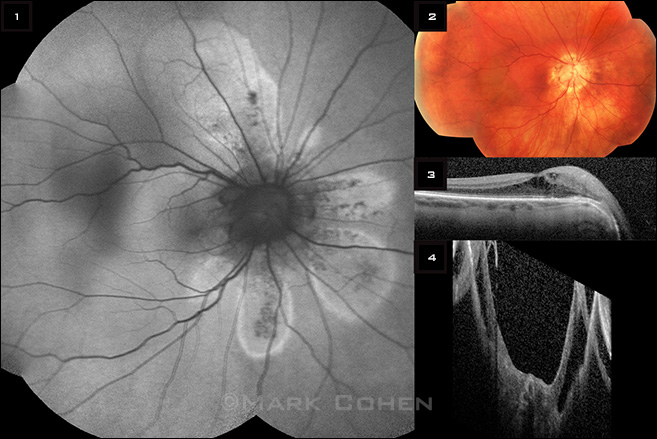
Fundus examination (Fig. 2) of the right eye revealed a large excavated optic disc with features of both optic pit and morning glory disc anomalies. There was loss of the foveal reflex, and subtle peripapillary pigmentary changes were visible.
Autofluorescence imaging (Fig. 1) demonstrated well-delineated areas of hyperfluorescence in a petalloid pattern. The hyperautofluorescent leading edge corresponded to the extent of the subretinal fluid. Optical coherence tomography (OCT; Fig. 3) showed that these areas corresponded to areas of serous retinal detachment and retinoschisis. Retinoschisis involving the fovea, seen on OCT (Fig. 4), was likely the cause of decreased visual acuity.
This case demonstrates the use of autofluorescence imaging in detecting (and, when appropriate, monitoring) serous retinal detachments and retinoschisis in patients with optic pit and morning glory disc anomalies.
| BLINK SUBMISSIONS: Send us your ophthalmic image and its explanation in 150-250 words. E-mail to eyenet@aao.org, fax to 415-561-8575, or mail to EyeNet Magazine, 655 Beach Street, San Francisco, CA 94109. Please note that EyeNet reserves the right to edit Blink submissions. |