This article is from November/December 2005 and may contain outdated material.
The patient had infectious uveitis—and she was surprisingly matter-of-fact about the underlying cause. “Of course I have syphilis,” she told Jean G. Deschenes, MD. “It is the fault of my husband.” Her response was uncommon, said Dr. Deschenes, professor of ophthalmology at McGill University in Montreal. “Usually, the patient jumps out of the chair.”
While most cases of uveitis are noninfectious and considered immune-mediated, a significant number are infectious in origin. According to Russell W. Read, MD, infectious uveitis (exclusive of cytomegalovirus retinitis) accounted for 13 percent to 21 percent of the total number of uveitis cases seen at tertiary referral centers.1 Dr. Read is associate professor of ophthalmology and pathology at the University of Alabama at Birmingham.
What’s the danger of underestimating this threat? Failing to give infectious etiologies a place on the diagnostic “radar screen” can have serious consequences for the patient, including loss of vision and, in some instances, a shortened life expectancy.
High Index of Suspicion
“Infections are with us forever, even if they are controlled,” said Khalid F. Tabbara, MD, professor of ophthalmology at King Saud University in Riyadh, Saudi Arabia. “Consider the viruses and tenacity of bacteria: These spirochetes have the talent of surviving for decades.”
Ancient diseases such as syphilis and tuberculosis still pose a threat, even as newer diseases like Lyme disease and West Nile have emerged. And air travel, immigration and globalization have overturned traditional patterns of geographic distribution of infectious diseases.
As a result, clinicians need to have a high index of suspicion, Dr. Deschenes said. When assessing a case of uveitis, “always rule out infectious uveitis first.”
 |
|
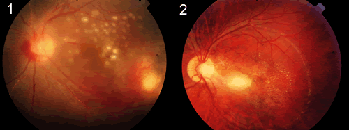
Uveitis With a Cause. (1) Multiple choroidal tuberculomas and (2) active toxoplasmic retinochoroiditis.
|
Potential Suspects
Here are some leading suspects to consider (see also the chart “Diagnoses Considered by Age”):
Cytomegalovirus. As of 2003, at least 405,926 people in the United States were living with AIDS.2 CMV-related retinitis is one of many opportunistic infections seen in HIV-infected patients with low CD4 cell counts (although it can also occur in heart or bone marrow transplant patients). Clinical findings include fine cell debris on the corneal endothelium, mild vitritis, and yellow-white slowly progressive retinitis occurring in any area of the retina, with retinal vasculitis and hemorrhages. Retinal detachment may occur.
Herpes. In addition to CMV, two other herpesviruses are common causes of infectious uveitis: herpes simplex and varicella zoster. In a study conducted in Los Angeles, 77 of 603 cases of uveitis (13 percent) were infectious in origin. Of those, 47 (61 percent) were due to herpes simplex and zoster. When considering anterior uveitis alone, every case was due to one of these two viruses.1 Herpes simplex uveitis may present with or without corneal disease. Other findings include moderate to marked vitritis, retinal hemorrhages, yellow-white peripheral necrotizing retinitis sparing the posterior pole, edema of the optic nerve head with retinal arteritis and retinal hemorrhages. Retinal detachment is common. Robert S. Weinberg, MD, noted that the presentation of herpes zoster can vary. “I’ve been fooled a couple of times” with patients in the prodromal stage, said Dr. Weinberg, associate professor of ophthalmology at the Wilmer Eye Institute.
Lyme disease. Borrelia burgdorferi infection is the most common vector-borne illness in the United States, with an incidence of 6.2 per 100,000 people, concentrated in northeastern and north-central states. Early findings include conjunctivitis and photophobia; later, Bell’s palsy and intermediate uveitis may occur.
Syphilis. Since 2000, this disease has increased 62 percent among men and 53 percent among women. In 2003, the incidence stood at 2.5 per 100,000 in the United States,2 with rates higher than that among gay men in urban centers.3 Syphilis is notoriously difficult to pin down—its nickname is “the great masquerader.” Clinical findings include anterior uveitis (either granulomatous or nongranulomatous), vitritis, retinitis and multifocal choroiditis. Iris roseola is a “classic sign,” said Dr. Weinberg.
Toxoplasmosis. Toxoplasmosis may be more common than previously thought. In a survey of 478 ophthalmologists, 55 percent indicated they had seen one or more patients thought to have active toxoplasmic retinochoroiditis in the previous two years, and 93 percent reported they had seen one or more patients with retinochoroidal scars thought to be inactive toxoplasmosis during that same time frame.,4Findings include yellow-white necrotizing retinitis with surrounding edema and a pigmented atrophic retinochoroiditic scar adjacent and contiguous to the lesion. Focal retinal vasculitis also may occur.
Tuberculosis. The overall incidence of TB in the United States is 5.1 per 100,000, and in those who were born abroad it’s higher—23.6 per 100,000.2 Findings include anterior granulomatous uveitis, mild to moderate vitritis and multifocal choroiditis.
Diagnoses Considered by Age
|
| < 4 |
Toxoplasmosis, CMV retinitis, syphilis, rubella |
| 5 to 15 |
Toxocariasis, toxoplasmosis, subacute sclerosing panencephalitis |
| 16 to 40 |
Lyme disease, candidiasis, toxoplasmosis, infectious mononucleosis |
| > 40 |
Herpetic necrotizing retinitis, tuberculosis, cryptococcosis, toxoplasmosis |
| AIDS (any age) |
CMV retinitis, herpetic retinitis, tuberculosis, toxoplasmosis, Mycobacterium avium complex infection, coccidioidomycosis, Pneumocystis carinii, candidiasis, aspergillosis. |
Source: Khalid F. Tabbara, MD
|
Diagnostic Challenges
Early diagnosis is imperative. Given the role that uveitis plays in blindness—an estimated 10 percent of all cases of blindness are due to the disease—there is no such thing as a benign ocular inflammation. “Think about diseases you can cure,” Dr. Weinberg emphasized.
Whether it’s herpes, TB or another etiology, the experts emphasized that you can’t rely on lab tests alone to pin down the diagnosis. Instead, the patient history is paramount.
“Whipple’s disease is a prime example of why we need to be compulsive with our patient history,” said Dr. Weinberg. “It’s relatively rare—but if you don’t think about it, you’ll miss it, and if you miss it, you’ll miss the chance to have a major impact on a patient’s life.”
And one patient interview may not be enough. “Be ready to revisit the history, over and over if needed,” said Dr. Read. The salient clues may emerge relatively late, during a second or third interview. For instance, has the patient recently spent time in the military— or in jail? Both involve overcrowding, which plays a role in TB transmission. More prosaically, does the patient have a cat? That might suggest toxoplasmosis or cat scratch disease.
Dr. Weinberg added another diagnostic tip: “Don’t think that you can make the diagnosis with pattern recognition. This is one of those times when that won’t work.” In addition, consider the natural history of the disease, the experts say. Then, once you have the critical clues in hand, you can do directed testing, Dr. Read noted.
In some cases, testing is of little value. The CMV antibody test, for instance, is not considered helpful, and the diagnosis of CMV retinitis is made on clinical grounds. Similarly, “we have seen many cases of active ocular TB without chest disease,” Dr. Tabbara cautioned.
________________________________
1 “Masquerading Infection: When Should You Suspect It?” Presented at the Academy’s Uveitis Subspecialty Day, Oct. 15, Chicago.
2 MMWR April 22, 2005;53:1–87.
3 MMWR July 9, 2004;53:575–578.
4 Lum, F. et al. Am J Ophthalmol 2005;140: 724.e1–724.e7.