By Samreen Khanam, MBBS, MS, Prolima Thacker, MBBS, MS, and Anju Rastogi, MBBS, MS
Edited By: Sharon Fekrat, MD, and Ingrid U. Scott, MD, MPH
Download PDF
Microspherophakia is a rare abnormality of the crystalline lens, marked by reduced equatorial diameter and increased lens thickness. The prevalence of this disorder is unknown, but it is reportedly more common in persons of Asian and North African descent.1
Although classically described in association with Weill-Marchesani syndrome (WMS), microspherophakia may occur with various ocular and systemic conditions, and in isolation.
Genetic Link to WMS
WMS is a rare inherited disorder of connective tissue, characterized by short stature, brachydactyly, brachycephaly, joint stiffness, maxillary hypoplasia, cardiovascular abnormalities, and ocular conditions. The latter include ectopia lentis, glaucoma, microspherophakia, and severe myopia.
Responsible mutations. Mutations in the ADAMTS10 and FBN1 genes have been identified in WMS. Another implicated mutation is that of the latent TGF-beta-binding protein 2 (LTBP2).1,2
ADAMTS10 mutation is known to cause the autosomal-recessive variant of WMS. The ADAMTS10 gene belongs to the ADAMTS family of zinc-dependent proteases associated with connective-tissue organization, coagulation, angiogenesis, and cell migration. These proteases play a major role in growth and in the development of the skin, heart, and crystalline lens.
The FBN1 gene encodes instructions for the protein fibrillin-1, which is an important component of connective tissue, giving it strength and flexibility. Mutations are inherited in an autosomal-dominant pattern and result in an unstable fibrillin-1, which weakens connective tissue and produces the syndrome’s ocular and systemic features.
Some cases of autosomal-recessive microspherophakia have been linked to mutations of the LTBP2.2 Structurally homologous to fibrillins, LTBPs are highly expressed in the lens capsule, lens epithelium, and ciliary body. ADAMTS17 is from the matrix metalloproteinase family of proteins that bind to the extracellular matrix and sequester TGF-beta. A number of mutations in the ADAMTS17 gene have been shown to disrupt the organization of the extracellular matrix, resulting in microspherophakia.1-3
 |
|
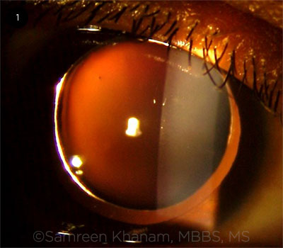
EDGE OF MICROSPHEROPHAKIC LENS. Pupil is fully dilated and the lens equator is visible. The patient had lenticular myopia of –11 D and features of angle-closure glaucoma. A clear lens extraction was performed, followed by in-the-bag IOL placement, which helped achieve better glaucoma control.
|
Pathophysiology
It has been observed that the human crystalline lens is almost spherical at birth, with an equatorial diameter of 6.5 mm and a sagittal diameter of 3.5 to 4.0 mm.4 As the child grows, the equatorial diameter increases rapidly, reaching about 9 to 10 mm by adulthood. The sagittal diameter is approximately 3.7 mm at 20 years of age and 4.0 mm at 50 years of age; the sagittal growth causes the lens to become more spherical with age.4
It has been postulated that weakness of the zonular fibers leads to lack of tension in the equatorial plane; thus, the lens remains spherical and does not acquire a biconvex shape.4 This spherical configuration of the lens results in a high degree of lenticular myopia in affected eyes. It also causes a shallow anterior chamber and, often, angle-closure glaucoma. Late subluxation of the lens may occur because of weak zonules.
Systemic and Ocular Links
Apart from WMS, microspherophakia has been associated with Alport syndrome, homocystinuria, Klinefelter syndrome, mandibulofacial dysostosis, and Marfan syndrome. It also has been linked to GEMSS syndrome (glaucoma, ectopia lentis, microspherophakia, stiff joints, and short stature). See Table 1 for more associations.
Familial microspherophakia is not associated with any systemic defect and is thought to be autosomal-recessive.
Clinical Features
Microspherophakia is usually bilateral. The edges of these small-diameter lenses generally can be observed when pupils are fully dilated (Fig. 1). Frequently, subluxation is present as well. Other common clinical observations are a high degree of lenticular myopia, defective accommodation, and angle-closure glaucoma.
Most patients who present to an ophthalmologist with microspherophakia will complain of low vision; however, some cases exhibit acute angle-closure glaucoma at initial presentation.5
Mechanisms of Glaucoma
Glaucoma associated with microspherophakia can result from various mechanisms. Pupillary block is a common mechanism leading to angle-closure glaucoma in patients with microspherophakia. Other reported mechanisms include crowding of the angle by the spherophakic lens, chronic pupillary block without complete angle closure, and angle abnormalities with agenesis of angle structures.
It is difficult to estimate the prevalence of glaucoma among patients with microspherophakia given the latter’s rarity. However, in a series of 36 eyes of patients with microspherophakia, glaucoma was present in 16 (44.4%).6
Examination
Ocular examination should include:
- Comprehensive slit-lamp exam: look at lens morphology and angle of the anterior chamber; look for preexisting subluxation
- Fundus exam: check for any glaucomatous damage to the disc
- Peripheral fundus exam: may rule out coexisting retinal pathology
- Intraocular pressure: measure, monitor, and manage appropriately
- Ultrasound biomicroscopy and anterior-segment optical coherence tomography: may help to elucidate biomechanics of angle crowding and angle closure in some patients
- Detailed systemic evaluation: mandatory to rule out syndromic association
- Measure lens thickness and axial length
Identifying Microspherophakia: Key Features
- Abnormally increased lens thickness
- Defective accommodation
- Features of angle closure
- Glaucomatous optic atrophy
- Isolated or syndromic association
- Lens edge seen in fully dilated pupils
- Lens subluxation or dislocation
- Low equatorial lens diameter
- Moderate or high myopia
- Shallow anterior chamber
|
Management
Lenticular myopia. Patients with microspherophakia who present with only lenticular myopia (without any other manifestation) need early refractive correction to avoid irreversible vision loss due to amblyopia. Routine follow-up will ensure that spectacle prescriptions are changed to coincide with the refractive status of the eye.
If glaucoma is present. Pupillary block can be relieved or prevented with Nd:YAG laser peripheral iridotomy. Cycloplegics are useful for managing acute attacks of pupillary block glaucoma. These drugs relax the ciliary muscle and tighten the zonules, in turn “pulling back” the iris-lens diaphragm, thus relieving the pupillary block.
If angle-closure glaucoma develops, it may require antiglaucoma medications. Eyes that do not respond to these medications may need glaucoma filtration surgery or a glaucoma drainage device. Primary trabeculectomy has a good success rate in such cases.7
As evidence mounts in support of early lensectomy in cases of primary angle closure, this approach could be considered in patients with microspherophakia, as well. However, it is important to note that recent studies looking at this issue did not include patients with secondary causes of angle closure.8
Lens subluxation. Clear lens extraction followed by in-the-bag intraocular lens (IOL) placement may relieve crowding of the angle by the spherophakic lens.9 However, this may be difficult to achieve in the presence of a small capsular bag and zonular instability. Therefore, capsular tension rings often are used in conjunction with the IOL. Other indications for lens extraction include subluxation, cataract, lenticulocorneal touch, and intermittent pupillary block.
Lensectomy by the limbal route is done in cases of severe lens subluxation and anterior dislocation.10 For posterior dislocation, a pars plana lensectomy is required.
Lifelong care. Most patients with microspherophakia will need lifelong ophthalmologic follow-up. It is important to make them aware of this.
___________________________
1 Bitar MS et al. JSM Ophthalmol. 2016;4(1):1040.
2 Kumar A et al. Hum Genet. 2010;128(4):365-371.
3 Morales J et al. Am J Hum Genet. 2009;85(5):558-568.
4 Chan RT, Collin HB. Clin Exp Optom. 2002;85(5):294-299.
5 Kaushik S et al. BMC Ophthalmol. 2006;6:29.
6 Muralidhar R et al. Eye. 2015;29(3):350-355.
7 Senthil S et al. Indian J Ophthalmol. 2014;62(5):601-605.
8 Azuara-Blanco A et al. Lancet. 2016;388:1389-1397.
9 Lu Y, Yang J. J Clin Exp Ophthalmol. 2016;7:532.
10 Rao DP et al. Br J Ophthalmol. 2018;102(6):790-795.
___________________________
Dr. Khanam is an ophthalmology resident and Dr. Rastogi is a Director Professor in the department of ophthalmology; both are at Guru Nanak Eye Centre in New Delhi. Dr. Thacker is an ophthalmologist in private practice in Lucknow, India. Financial disclosures: None.