Download PDF
Arnav Bhutia* was an active 5-year-old who enjoyed wrestling with his older brothers. When his parents noticed that Arnav’s right eye looked swollen, they initially assumed that it had occurred during horseplay.
When the swelling hadn’t improved after a few days, they gave him an oral antihistamine and topical Zaditor (ketotifen fumarate) allergy drops, but there was no change.
One week later, when Arnav was seen by the family’s pediatrician, the right eye was partly closed, and the right upper lid looked swollen to the doctor. But Arnav had no pain or itching, and neither the eye itself nor the lid was red. With a history of sinusitis in the past, Arnav was put on oral cefdinir and oral prednisone.
However, Arnav’s eye continued to swell for the next three days. By now, it was the weekend, and his increasingly concerned parents took him to the local emergency department (ED). Because Arnav had experienced a tick bite in the past, the ED physician ordered a Lyme serology test; it returned negative. A complete blood count was also normal. No imaging studies were performed in the ED. Arnav’s antibiotic prescription was changed to oral ciprofloxacin.
The next day, Arnav began noticing double vision and was referred to ophthalmology by the pediatrician.
We Get a Look
Arnav was a friendly, smiling child with significant ptosis of the right upper lid, resulting in a 2-mm palpebral fissure. There was no redness, but he had proptosis measuring 2 mm on Hertel exophthalmometry and a definite upgaze restriction in the right eye. The left eye appeared normal on external exam. Vision was 20/30 in the right eye and 20/25 in the left eye; in both eyes, pupil reactions were normal, with no afferent pupillary defect. On dilated exam, optic nerves and retinas appeared normal. Given Arnav’s rapidly worsening appearance, along with proptosis, diplopia, and ptosis, an immediate computed tomography (CT) scan was performed.
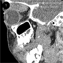
The CT scan showed an enhancing extraconal soft-tissue mass, measuring 3.2 × 2.8 × 1.6 cm, along the superior aspect of the right orbit with no bony erosion (Fig. 1). After the discovery of this orbital mass, the patient was sent for immediate biopsy by an oculoplastic surgeon.
 |
|
WHAT IS IT? Sagittal CT scan of our patient shows a solid tumor in the superior orbit pushing the globe downward. There is no bony erosion.
|
Differential Diagnosis
For a pediatric patient presenting with proptosis, severe ptosis, and diplopia, the differential diagnosis divides into four categories: infection, trauma, inflammation, and tumor. There are multiple possible conditions within these groups.
Infection. Arnav’s condition was initially misdiagnosed as orbital cellulitis because of the ptosis and rapid progression. Typically, infection spreads from the paranasal sinus to the orbit, but it can also originate from other adjacent sinuses, the skin, or the bloodstream. A patient with orbital cellulitis typically presents not only with ptosis and proptosis but also with common symptoms of infection, including fever and leukocytosis. A CT scan demonstrating inflamed extraocular muscles can confirm the diagnosis.
Treatment involves immediate administration of broad-spectrum intravenous antibiotics, followed by collecting and culturing specimens to determine the causative organism. An abscess may require surgical drainage.
Because Arnav had no pain, fever or leukocytosis, we eliminated orbital cellulitis from our differential.
Trauma. Although orbital trauma is included in the differential, we would expect there to be significant pain as well as erythema and bruising if Arnav had been hit hard enough to cause nearly complete ptosis. Orbital hemorrhage, which would have shown a characteristic appearance on CT or magnetic resonance imaging (MRI), can cause proptosis, and direct damage to the levator muscle can contribute to ptosis.1
The treatment varies depending on the injury. Some cases of orbital trauma heal spontaneously, while others require surgical intervention, as indicated by the presence of significant orbital hemorrhage or permanent damage to levator function.
Inflammation. Orbital inflammatory syndrome, also known as orbital pseudotumor, involves invasion of the orbit by plasma cells, lymphocytes, eosinophils, and fibroblasts. The cause is unknown, and patients typically experience an abrupt onset of pain, swelling, diplopia, and proptosis. Diagnosis can usually be made with CT or MRI but occasionally requires a biopsy due to the nonspecific appearance of orbital edema.
Corticosteroids are used to treat the inflammation, and rapid improvement is expected within 24 to 48 hours of administration.
Tumors. Proptosis is a classic presentation for an orbital tumor. Suspected tumors must be addressed swiftly, as survival hinges on rapid treatment for certain entities in this category. The differential includes vascular tumors as well as lymphomas, leukemias, neuroblastomas, and rhabdomyosarcomas.
Vascular. In children, vascular tumors are among the more common tumor subcategories, with capillary hemangiomas and lymphangiomas being the most frequent.2 These two are benign and are typically found in very young children. Symptoms present slowly over the course of months to years, in contrast to Arnav’s experience.
With lymphangiomas, mild trauma can cause hemorrhage into the periorbital area, which could result in a presentation much like Arnav’s. However, we eliminated this possibility because lymphangiomas have a specific cystic appearance on CT that was absent on our patient’s scan.
Another type of vascular tumor, cavernous hemangioma, is found in adults, grows relatively slowly, and has a characteristic radiologic appearance not seen in our patient.
Malignant. Orbital Hodgkin lymphoma is a rare childhood tumor presenting most commonly in adolescents, is often associated with immunodeficiencies, and can be familial. The patient usually has fatigue, fever, and weight loss in addition to ocular findings.
Another malignant orbital tumor is orbital granulocytic sarcoma, a rare variant of acute myelogenous leukemia. It presents over the course of one to two months with proptosis and fever. It is typically diagnosed histologically through fine-needle aspiration biopsy. After diagnosis, chemotherapy is the preferred treatment.3
Neuroblastoma is the most common cause of metastasis to the orbit in childhood. These tumors typically present with bruising around the eye and systemic symptoms such as pain and weakness.
Orbital rhabdomyosarcomas are tumors of the orbit that develop from striated muscles. The tumor is confirmed through various histological analyses and immunoreactive stains.
Intraoperative Findings
The plan was to do an incisional biopsy. However, after making an incision of approximately 4 cm in the upper eyelid and locating the lesion, the surgeon determined that the tumor had a discrete capsule and could be removed in its entirety, which measured 4 × 5 × 3 cm.
Histology showed a spindle-cell neoplasm with mildly pleomorphic cells within a myoid background. There were frequent mitoses. Many of the cells demonstrated nuclear staining with MyD1, and a number of cells were immunoreactive for myogenin and Myf4. The neoplasm was positive for vimentin. The cells were negative for S100, desmin, CD45RB, and smooth muscle actin. Flow cytometry was negative for non-Hodgkin lymphoma. Testing was also negative for 13q14.1 (FOX01) rearrangement. Histological analysis of the sample confirmed that the tumor was consistent with embryonal rhabdomyosarcoma.
Additional workup was performed for staging purposes. It included CT scans of the nasopharynx, neck, chest, and pelvis, which were found to be negative for metastasis.
About the Disease
Orbital rhabdomyosarcoma is the most common primary orbital malignant tumor in childhood. Overall, rhabdomyosarcoma has an incidence of 4.3 cases per 1,000,000, with 10% of these cases occurring in the orbit. Orbital rhabdomyosarcoma is most often found in patients under 16 years old (mean age of onset is between 5 and 7 years).4 It is usually painless (approximately 10% of patients report pain) and presents rapidly with proptosis (about 80%), globe displacement (80%), lid swelling (60%), and ptosis (30%-50%).5
Orbital rhabdomyosarcoma should be considered in the differential diagnosis of almost all pediatric patients presenting with ptosis or proptosis. Diagnosis can be confirmed with CT or MRI. On a CT scan, orbital rhabdomyosarcoma appears as a homogeneous, isodense soft-tissue mass with or without bone destruction (depending on the stage of the cancer).5
Clinical groups. The North American Intergroup Rhabdomyosarcoma Study Group (IRSG) has defined four groups of orbital rhabdomyosarcoma. This grouping is based on the results of the excisional surgery and provides guidelines for postsurgical care of the cancer. Our patient had localized disease and underwent complete surgical excision of the tumor, placing him in Group I. Postoperative treatment for a patient with Group I orbital rhabdomyosarcoma is a chemotherapy regimen, typically vincristine, dactinomycin, and cyclophosphamide, for eight cycles (24 weeks). Radiation is not used in these cases. However, patients in Groups II, III, and IV are treated with both chemotherapy and radiation ranging from 36 Gy to 46 Gy.
Prognosis. Compared to Group I, the prognosis for Groups II to IV is worse, with a five-year survival rate ranging from 75% to 87%.
Group I orbital rhabdomyosarcoma prognoses are generally quite favorable, but the results vary depending on the pathological origin. There are three subtypes, two of which are found in children (embryonal and alveolar). These two can be differentiated through cytogenetic and histological analysis.
Alveolar rhabdomyosarcoma is less frequent (approximately 10% of cases versus 65% for embryonal, with the remaining percentage divided between spindle cell, botryoid, and undifferentiated variants) and more aggressive than embryonal rhabdomyosarcoma. The alveolar type gets its name from its histological appearance, which is similar to lung alveoli. Most alveolar orbital rhabdomyosarcomas (80%) have a genetic translocation involving the PAX and FOX01 genes, which promotes the lack of differentiation and increased proliferation found in the tumor cells. A fluorescence in situ hybridization analysis can be used to identify a translocation; in Arnav’s case, the results of this test were negative.
The histopathological analysis confirmed an embryonal orbital rhabdomyosarcoma in our patient. The prognosis for Group I is highly favorable (94% five-year survival rate).6 Embryonal rhabdomyosarcoma stems from the proliferation of myogenic precursor cells, which are under the control of transcription factors such as myogenin and Myf4. Histological staining showed some cells carrying these two transcription factors, confirming the diagnosis of an embryonal rhabdomyosarcoma.
Grand Rounds
Don’t miss this year’s Grand Rounds symposium on Sunday Nov. 15, 2:05-2:50 p.m. PST, with residents presenting real cases to a panel of experts.
|
Our Patient’s Progress
In addition to surgical excision, Arnav received chemotherapy with vincristine, dactinomycin, and cyclophosphamide. Subsequent MRIs at one and two years have been negative. Other than having a gradually fading scar on his upper lid, Arnav is a healthy, active child who continues to enjoy wrestling with his older brothers.
___________________________
1 Jacobs SM et al. J Ophthalmic Vis Res. 2018;13(4):447-452.
2 Castillo BV Jr, Kaufman L. Pediatr Clin North Am. 2003;50(1):149-172.
3 Thakur B et al. J Clin Diagn Res. 2013;7(8):1704-1706.
4 Wharam M et al. Ophthalmology. 1987;94(3):251-254.
5 Shields JA, Shields CL. Surv Ophthalmol. 2003;48(1):39-57.
6 Tang LY et al. Cancer Manag Res. 2018;10:1727-1734.
___________________________
*Patient’s name is fictitious.
___________________________
Mr. Metzman is a second-year medical student at the Indiana University School of Medicine in South Bend. Dr. Gerber is in practice at Advanced Ophthalmology and is a clinical professor of ophthalmology at the Indiana University School of Medicine in South Bend. Financial disclosures: None.