Download PDF
Can spectral-domain optical coherence tomography (SD-OCT) help clinicians detect structural glaucomatous damage and the changes associated with the diagnosis of glaucoma? Yes and yes, according to an Academy Ophthalmic Technology Assessment (OTA).1
“Classic structural changes associated with glaucoma can be detected in the retinal nerve fiber layer, the macula, and the optic nerve with SD-OCT technology,” said Teresa C. Chen, MD, at Harvard Medical School in Boston. She called SD-OCT “a useful tool in the management of glaucoma patients.”
Expansion in knowledge. The literature review began where the previous imaging OTA left off—February 2006—and concluded in April 2018. During that time, 708 articles on the use of SD-OCT to help clinicians detect changes in eyes diagnosed with glaucoma appeared in the literature. Of those, 74 met inclusion criteria, with 2 identified as level I, and 57 as level II. The remaining 15 articles were not used in the analysis.
 |
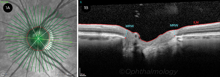
CONFIRMATION. Radial scan (1A) and corresponding SD-OCT image (1B). In this imaging example, SD-OCT was used to rule out glaucoma in myopic eyes. MRW = minimum rim width; ILM = internal limiting membrane.
|
Expansion in technology. “Most clinical practices have transitioned from the older 2-D time-domain OCT machines to the newer 3-D SD-OCT machines,” Dr. Chen said. In the studies evaluated in the OTA, the Cirrus High-Definition OCT (Carl Zeiss Meditec) was the most commonly studied machine, followed by the RTVue-100 (Optovue), the Spectralis SD-OCT (Heidelberg Engineering), and the 3D OCT-1000 and 3D OCT-2000 (Topcon).
Results. “Though different machines have different scan protocols and different software packages, all can detect the same classic pattern of structural changes noted in glaucoma—superior and inferior thinning,” said Dr. Chen. Findings from the OTA include the following:
- All instruments were capable of detecting damage to the retinal nerve fiber layer (RNFL), macula, and optic nerve in patients with preperimetric and perimetric glaucoma.
- RNFL was the most commonly studied single parameter, followed by the macula and optic nerve.
- All instruments can detect the same typical pattern of glaucomatous RNFL loss that affects primarily the inferior, inferior temporal, superior, and superior temporal regions of the optic nerve.
- The best disc parameters for detecting glaucomatous nerve damage are global rim area, inferior rim area, and vertical cup-to-disc ratio.
- Newer reference-plane independent optic nerve parameters may have the same or better detection capability when compared with older reference-plane dependent disc parameters.
Bottom line. The OTA does caution clinicians to be aware of factors that may influence test results, including “testing artifacts, false positives, false negatives, refractive error … and normal aging changes.” But overall, “SD-OCT machines allow for better axial resolutions, faster acquisition speeds, better scan quality, and better reproducibility, all of which affords us better information to care for our patients,” Dr. Chen said.
—Miriam Karmel
___________________________
1 Chen TC et al. Ophthalmology. 2018;125(11):1817-1827.
___________________________
Relevant financial disclosures—Dr. Chen: None.
For full disclosures and the disclosure key, see below.
Full Financial Disclosures
Dr. Chen U.S. Department of Defense: S; Harvard Foundation Grant (Fidelity Charitable Fund): S.
Dr. Drenser Interview Medical Systems: O,P; Phoenix: O; Retinal Solutions: O,P; Spark Therapeutics: C.
Prof. Dua Croma: C; Dompé: C; GlaxoSmithKline: O; NuVision Biotherapeutic: O; Santen: C; Shire: C; Thea: C; VisuFarma: C.
Dr. Kelly U.S. Department of Defense: S; Neuro Kinetics: S.
Disclosure Category
|
Code
|
Description
|
| Consultant/Advisor |
C |
Consultant fee, paid advisory boards, or fees for attending a meeting. |
| Employee |
E |
Employed by a commercial company. |
| Speakers bureau |
L |
Lecture fees or honoraria, travel fees or reimbursements when speaking at the invitation of a commercial company. |
| Equity owner |
O |
Equity ownership/stock options in publicly or privately traded firms, excluding mutual funds. |
| Patents/Royalty |
P |
Patents and/or royalties for intellectual property. |
| Grant support |
S |
Grant support or other financial support to the investigator from all sources, including research support from government agencies (e.g., NIH), foundations, device manufacturers, and/or pharmaceutical companies. |
|
More from this month’s News in Review