Download PDF
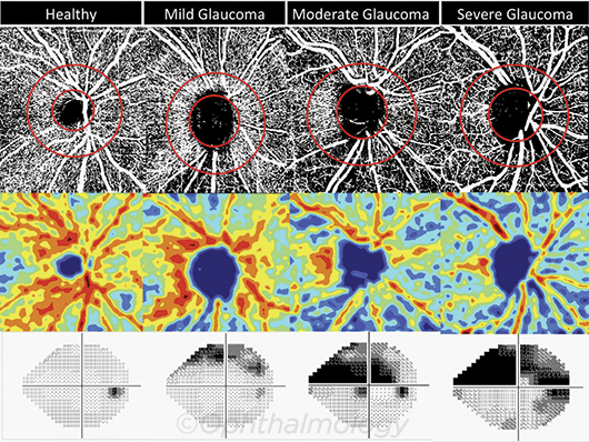
Using advanced imaging technology, researchers at the University of California, San Diego, have found a significant relationship between vessel density and severity of visual field (VF) damage in glaucoma.1 The microvascular network got progressively sparser as disease advanced, suggesting that reduced vessel density is associated with more severe disease.
The observational cross-sectional study involved 153 eyes from healthy participants, glaucoma suspects, and glaucoma patients. All eyes underwent imaging using optical coherence tomography angiography (OCT-A) and spectral domain OCT (SD-OCT); VFs were checked with standard automated perimetry. It was OCT-A that yielded information with the potential to transform clinical practice and provide a deeper understanding of the pathophysiology of glaucoma.
 |
VESSELS AND VF IN HEALTHY AND AFFECTED EYES. (Top row) OCT-A vessel density extracted maps; circles show measurement region. (Middle) Color-coded maps of vessel density. (Bottom) Corresponding visual fields.
|
OCT-A reveals RNFL capillary networks. OCT-A provides a window into the microvasculature in the optic nerve head, peripapillary retina, and macula, revealing ocular circulation “at a level of precision not achieved with previous instruments,” the researchers noted.
They evaluated vessel density, which they defined as the percentage of area occupied by flowing blood vessels in 2 designated regions of the retinal nerve fiber layer (RNFL). After comparing OCT-A observations to standard SD-OCT structural measures, the researchers found that healthy eyes appeared to have denser capillary networks within the RNFL than eyes with glaucoma. Vessel density was lowest in eyes with the most severe disease.
Linkage with VF damage. Even after controlling for the severity of structural damage, as measured by rim area and RNFL thickness, the association between vessel density and severity of visual field damage was significant. That strong correlation was an unexpected finding, said Robert N. Weinreb, MD, chairman and distinguished professor of ophthalmology, UC San Diego, who is one of the study’s authors.
Dr. Weinreb noted that these findings must be confirmed by others, and information regarding changes over time is needed “before employing this promising new technology for clinical decision making in glaucoma.” Eventually, he said, “It may be that these measurements will enhance our understanding of the pathophysiology of the disease and, specifically, the role of microvascular changes.”
—Miriam Karmel
___________________________
1 Yarmohammadi A et al. Ophthalmology. 2016;123(12):2498-2508.
___________________________
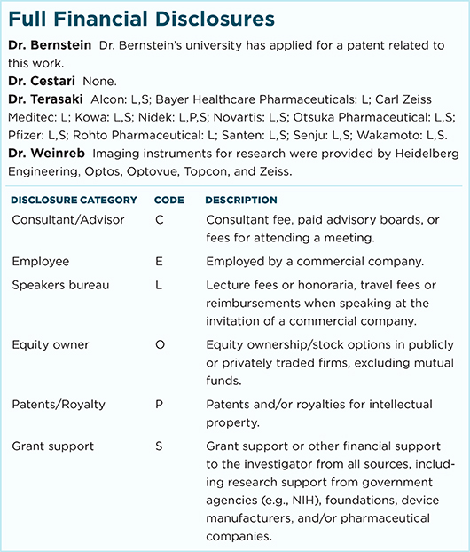
Relevant financial disclosures—Dr. Weinreb: Imaging instruments for research were provided by Heidelberg, Optos, Optovue, Topcon, and Zeiss.
For full disclosures and disclosure key, see below.

More from this month’s News in Review