Blink
FEB 01, 2019
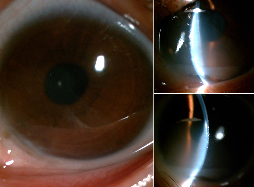
Can You Guess February's Mystery Condition?
Download PDF
Make your diagnosis in the comments, and look for the answer in next month’s Blink.

Last Month’s Blink
Presumed Ocular Histoplasmosis Syndrome
Written by Philip L. Ames, MD, Kathleen A. Regan, MD, and Siva S. Radhakrishnan Iyer, MD. Photos by Dr. Iyer, University of Florida College of Medicine, Gainesville, Fla.
A 62-year-old woman presented withblurry vision in her right eye, which had started 3 weeks prior to her visit. Her ocular history was significant for long-standing poor vision in the left eye from presumed ocular histoplasmosis syndrome (POHS). On exam, the best-corrected visual acuity was 20/60 in her right eye and count fingers at 1 foot in her left. Intraocular pressure was normal in both eyes.
Dilated funduscopic exam of the right eye (Fig. 1) showed a 1/4 disc–diameter gray lesion in the inferior macula with subretinal fluid and associated small hemorrhage. Both eyes had peripapillary atrophy with punched-out lesions in the midperiphery. In the left eye, disciform macular scarring was present (Fig. 2). Fluorescein angiography of the right eye (Fig. 3) illustrated early staining with late leakage in the inferior macula consistent with a choroidal neovascular membrane (CNVM). Optical coherence tomography of the right eye (Fig. 4) confirmed ellipsoid zone disruption from a subretinal lesion with associated fluid.
Intravitreal bevacizumab treatment was initiated in the right eye only, with involution of the CNVM and complete resolution of subretinal fluid after 3 treatments. Vision in this eye improved to 20/20.
POHS may develop a CNVM with an annual incidence of 1.8%.1 These lesions are very responsive to anti-VEGF as in this case.
___________________________
1 Macular Photocoagulation Study Group. Arch Ophthalmol. 1996;114(6):677-688.
Read your colleagues’ discussion.
| BLINK SUBMISSIONS: Send us your ophthalmic image and its explanation in 150-250 words. E-mail to eyenet@aao.org, fax to 415-561-8575, or mail to EyeNet Magazine, 655 Beach Street, San Francisco, CA 94109. Please note that EyeNet reserves the right to edit Blink submissions. |