Download PDF
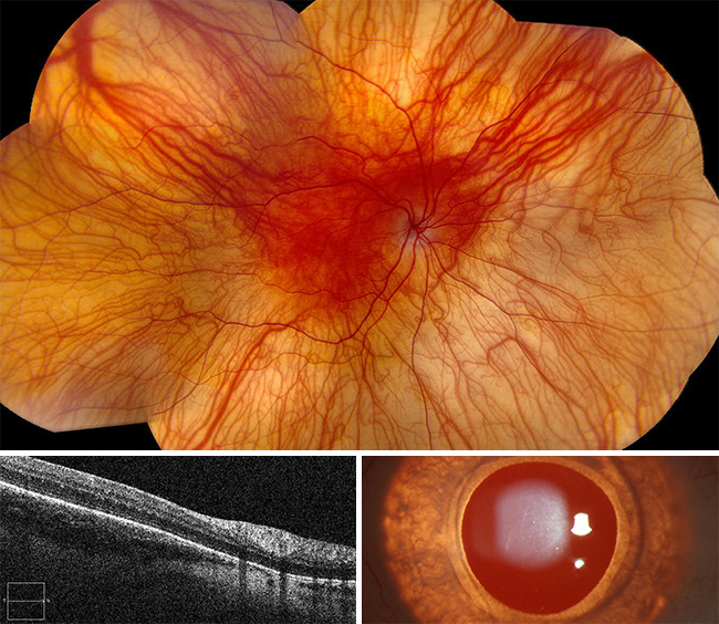
Make your diagnosis in the comments, and look for the answer in next month’s Blink.

Last Month’s Blink
Posterior Scleritis
Written by Alec Chaleff, MD, MBA, Hershel Rajendrakumar Patel, MD, MS, and Peter R. Pavan, MD. Photo by Peter R. Pavan, MD. All are at the University of South Florida Eye Institute, Morsani College of Medicine, Tampa, Fla.
A 68-year-old Hispanic woman presented with pain behind her right eye. She reported a shadow inferiorly in her right field of vision and flashing lights in her right eye after waking up. Visual acuity was 20/150 OD and 20/50 OS with normal intraocular pressure OU. Confrontation visual field revealed an inferior nasal and temporal defect OD.
Slit-lamp examination showed injected conjunctiva and nuclear sclerosis bilaterally. Funduscopy revealed Weiss rings in the vitreous OU. There was an elevated, slightly pigmented mass OD 1 disc diameter superior to the nerve, extending anteriorly and temporally. It was at least 3 × 3 (disc diameters) with possible subretinal fluid present anteriorly (Fig. 1).
Color photography showed that the mass was elevated and chorioretinal folds were present along the posterior edges. B-scan OD revealed a positive T-sign with fluid in Tenon space (Fig. 2) and neurosensory detachment inferior to the mass. The mass had low reflectivity over the anterior half, with the posterior half demonstrating higher reflectivity. Retro-orbital low reflectivity around the superior rectus muscle was also noted (Fig. 3). Fluorescein angiography (FA) OD revealed staining over the lesion with minimal late leakage. There was no intrinsic vascularity (Fig. 4).
After an autoimmune and infectious workup, the patient was diagnosed with posterior scleritis, with fluid in Tenon space. She was started on indomethacin and asked to return in 1 month.
At follow-up, the ocular pain had resolved, and her visual acuity was 20/60 OD and 20/40 OS. The mass was virtually flat and smaller, and chorioretinal folds were no longer present. B-scan showed high to medium reflectivity, and marked leakage was no longer present.
Read your colleagues’ discussion.
| BLINK SUBMISSIONS: Send us your ophthalmic image and its explanation in 150-250 words. E-mail to eyenet@aao.org, fax to 415-561-8575, or mail to EyeNet Magazine, 655 Beach Street, San Francisco, CA 94109. Please note that EyeNet reserves the right to edit Blink submissions. |