Download PDF
Researchers have known for almost a century that our fovea has a much lower sensitivity to rapidly changing visual inputs than our peripheral vision. This lesser sensitivity to rapid changes in visual stimuli is what enables humans to perceive continuous motion when we focus our central gaze on images in a flipbook, a movie, or a TV show.
Past work has shown that this perceptual difference might originate in the retina rather than in the higher brain centers. But the question has remained: Where in the retinal circuit does this originate, and what are the underlying neural mechanisms?
 |
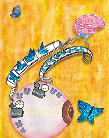
SPEED OF PROCESSING. In this schematic illustration, visual information encoding the flight of a butterfly is conveyed by cone photoreceptors depicted as movie cameras with clocks that have different gradations. The “foveal camera clock” has coarser gradations, and the “film reel” carrying the information of the butterfly’s flight to the brain with the highest spatial resolution has a slower “frame rate,” with half as many frames compared with the peripheral film reel.
|
Intracellular recordings provide the clues. Scientists have now determined that cone photoreceptors in the fovea process incoming light in a different way from those in the peripheral retina—and that accounts for the difference in the temporal sensitivity of retinal signals sent to the brain. The discovery, based on recordings from individual neurons in the primate fovea, was reported by researchers at the University of Washington.1
“We did the first intracellular recordings from cone photoreceptors and the retinal output neurons, the ganglion cells, and systematically compared the electrical responses in them with their counterparts in the peripheral retina. And what we found is that this difference in temporal sensitivity in our visual perception is present in the cone photoreceptors themselves,” said coauthor Raunak Sinha, PhD, a research fellow in the Department of Physiology and Biophysics at the University of Washington School of Medicine, in Seattle.
“We were further able to trace the reason down to the cellular mechanism,” Dr. Sinha said. “The phototransduction cascade—the very first stage of our visual processing—is essentially different between foveal and peripheral cone photoreceptors. That was a big surprise.”
Tracking the signals. These observations were made in vitro, using monkey retinas that continue to function normally for about 24 hours in the laboratory, Dr. Sinha said. The researchers were able to measure both the amount of light entering the individual photoreceptors and the output signals that they and cells downstream in the neural circuit emitted.
These measurements confirmed that peripheral cones transduce incoming light into electrical signals, which then flow sequentially to bipolar cells and retinal ganglion cells (RGCs) on their way to the visual cortex, he said. As they pass through the neural circuits to the brain, the signals are regulated at the synapses through the well-known mechanism of neural inhibition, he said.
But, in the fovea, molecular tests to detect inhibitory receptors on dendrites of ganglion cells showed that the dominant neural circuit—the midget ganglion cells—operates effectively independent of conventional synaptic inhibition. This is contrary to other RGCs, including peripheral midget ganglion cells, Dr. Sinha said.
This difference was a key insight for the researchers, but it does not account for differences in temporal sensitivity between the 2 retinal regions, he said.
Instead, the electrical measurements showed that phototransduction in the cones exhibits a 2-fold difference in response time, which is nearly identical to the difference in perceptual sensitivity between foveal and peripheral vision. “Our results provide a simple explanation for a salient perceptual observation,” Dr. Sinha said.
How does this help? Going forward, the techniques used in the study will be important for researchers seeking to better understand diseases that perturb foveal signaling, such as macular degeneration, Dr. Sinha said. Furthermore, knowing that there is a fundamental difference in the computational strategies employed by foveal and peripheral retina will help shape the algorithms that scientists use to design visual prostheses to better mimic human vision, he said.
—Linda Roach
___________________________
1 Sinha R et al. Cell. 2017;168(3):413-426.
___________________________
Relevant financial interests—Dr. Sinha: None.
For full disclosures and disclosure key, see below.

More from this month’s News in Review