When I explain to parents, residents and colleagues why I chose pediatric ophthalmology as a subspecialty, I always include in my reasons, “I get to play all day long.” I use toys, funny faces, weird noises, beat boxing, singing and play to get the examination I need from children.
Conquering the pediatric exam takes several keys: be observant, be quick, be flexible, be fun. In addition, know the goal of your examination. What is the chief complaint? What is the most important part of your entire examination?
Consider starting with that last question, since children have short attention spans. For difficult patients, work from least confrontational (corneal light reflex and motility) to most “in your face” (alternating cover test, monocular visual acuity).
In the era of cameras in every parent’s pocket, don’t hesitate to ask the parents for examples of what they are seeing at home.
Here’s how to conquer each part of your pediatric examination.
Motility and alignment:
 You can use anything to assess motility (extraocular muscle function). This includes lighted toys, your phone tuned to YouTube (Little Baby Bum), Netflix (Moana) or your face (best for infants). In general, you get one look per toy. Have a pocket or drawer filled with fun, visually interesting toys.
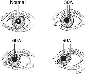
When measuring alignment, kids can be scared of occluders. As an alternate, your thumb or hand can serve as an excellent occluder. You can also use a Hirschberg light reflex (cornea light reflex with a Finoff light) for children who won’t let you get near them. Light reflex outside the pupil = esotropia; inside pupil = exotropia.
Visual acuity:
Fix/follow only provides a rudimentary idea of a patient’s vision. Do not use a toy that makes any noise, as you may unintentionally test hearing instead of vision.
When testing subjective acuity, do not be afraid to patch the contralateral eye. Children are excellent peekers. Do not trust them! I often test vision with Allen (pictures), HOTV eye chart (using crowding bars if testing with single letters as you will overestimate vision without them) or Snellen chart.
If the child cannot participate in subjective acuity, a central/steady/maintained (CSM) approach will give you excellent information about fixation preferences and presence of amblyopia.
- Central (monocular test): review central or eccentric fixation.
- Steady (monocular test): look for absence or presence of nystagmus or roving eye movements.
- Maintained (binocular test): see if the child can hold fixation with the eye. For children with eye misalignment, can the patient maintain fixation with the viewing eye when you uncover the previously covered eye?
If all else fails, equal versus unequal objection to occlusion (for example, crying when one eye is patched but not the other) can give excellent information.
Pupils:
The retinoscope provides an excellent way to check for anisocoria from a distance. Place the retinoscope light bar at 180 degrees, sit far enough from the child to have the light hover over both pupils and peer into the retinoscope. This is good for dim and light settings, as well as dark irises.
Confrontational visual fields:
Use the two-toy method: have a child fix on a toy in front of them. Slowly bring a second toy into the child’s visual field. If they then look toward the new toy (either with their entire head or their eyes), then they can see in that field!
Globe examination:
The red reflex test with a retinoscope can give you excellent information about the state of a child’s cornea, iris shape and lens without having the child sit at a slit lamp. Do this on every child and you will pick up small opacities you may otherwise have missed.
Fundus:
Good luck. Distraction is key. Use your phone again for videos. Ask the child what animals they think you’ll find in their eyes. Make sure you get a good look at the macula and optic nerves. The 28 diopter lens allows you get see more than a 20 diopter lens (70° vs 60°), but you sacrifice zoom (2.27× vs 3.13×).
Refraction:
Cycloplegic retinoscopy is key for every child’s examination. Watch this excellent tutorial to understand how retinoscopy works. Expert tips:
- If your reflex already appears neutralized, but blunted, without any lenses, the child likely has a high refractive error. Try a –10.00 and a +10.00 and see what happens.
- It’s much easier to see “with” motion of the retinoscope reflex than “against” motion. I prefer to work my way up from less plus (or more minus) to the neutralized reflex.
- Know your working distance. Don’t forget to subtract 1/working distance (in meters) from the lens you used to neutralize the reflex. Measure your working distance on day 1.
Bottom line: Kids are fun. Have fun with them and trick them into giving you the crucial information you need to help them achieve a lifetime of good vision.
* * *
 About the author: Evan Silverstein, MD, is an assistant professor of ophthalmology and associate residency program director at Virginia Commonwealth University in Richmond, Va. Dr. Silverstein completed his residency at Vanderbilt University and a pediatric and adult strabismus fellowship at Duke University. He joined the YO Info editorial board in 2017.