40-year-old man with blurred vision, pain, redness, and tearing in his left eye.
Cataract/Anterior Segment
What is your diagnosis?
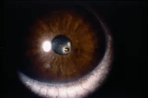
The diagnosis is...
![]()

The image is consistent with a diagnosis of corneal foreign body.
- Metallic fragments, wood splinters, and fiberglass can all potentially become foreign bodies.
- Risk factors include engagement in high-risk activities such as welding, hammering, and drilling. Use of protective eye wear can greatly reduce the risk.
- A foreign body can lodge in any layer of the cornea, potentially leading to an open-globe injury.
What is the role of the primary care or emergency medicine physician?
![]()
Emergently refer the patient to an ophthalmologist for evaluation of an open-globe injury and other complications, including reactive iritis.
What is the role of the ophthalmologist?
![]()
- An ophthalmologist will identify the foreign body and evaluate the extent of the damage under a slit lamp, utilizing certain dyes (eg, fluorescein).
- If a globe perforation is identified, a rigid eye shield should be placed over the eye to prevent extrusion of intraocular contents.
What is the treatment?
![]()
- The removal method depends upon the depth of impaction of the foreign body and may require surgery.
- If the object is metallic, the rust ring must be removed to prevent further irritation and infection.
- Topical antibiotics and a protective soft contact lens may be applied after removal of the foreign body.
- Treatment will also depend on whether soil or other debris came in contact with the eye.
Learn more: Ophthalmology resources for medical students
![]()