By Ahmadreza Moradi, MD, and Rona Z. Silkiss, MD, FACS
Edited By: Ingrid U. Scott, MD, MPH
Download PDF
Throughout most of his adult life, Joe Smith* was used to hearing the same refrain: “You look sleepy.” Furthermore, over the previous decade, the 57-year-old had been receiving that comment more and more often. He also had been experiencing increasing difficulty with driving, reading, and using the computer, even though he hadn’t noticed any vision changes or diplopia. His optometrist diagnosed ptosis in both eyes, and he was referred to our clinic for evaluation.
We Get a Look
History. Mr. Smith was diagnosed with hypertension in 2009 for which he was prescribed lisinopril and amlodipine. His blood pressure was well controlled, and he had no other significant past medical history. His mother and brother have a similar “sleepy” facial appearance.
Exam. On examination, his best-corrected visual acuity was 20/20 in each eye. His intraocular pressure was 14 mm Hg in the right eye and 13 mm Hg in the left. Visual fields were diminished bilaterally. There was no afferent pupillary defect. Color vision was intact in both eyes. Ocular motility was restricted in both eyes. The margin to reflex distance (MRD1) was –1 bilaterally. He also had head tilt.
The slit-lamp examination showed a deep and quiet anterior chamber in each eye. The dilated examination was unremarkable in both eyes.
Differential diagnosis. Clinically, Mr. Smith presented with bilateral progressive ptosis with ophthalmoplegia. Our differential diagnosis was broad and included acquired ptosis (such as aponeurotic ptosis, postsurgical ptosis, posttraumatic ptosis), myasthenia gravis, congenital ptosis, Kearns-Sayre syndrome, oculopharyngeal musculodystrophy, and chronic progressive external ophthalmoplegia (CPEO).
Mr. Smith did not complain of problems with swallowing, hearing loss, or nonocular muscle weakness. He hadn’t undergone any ocular surgery, and he had not experienced trauma or any cardiovascular symptoms.
Definitive diagnosis. The findings on examination of ptosis and extraocular muscle restriction (Fig. 1) are characteristic of isolated CPEO. This was reinforced by the facial appearance of Mr. Smith’s mother and brother, which suggested that the family history is positive for CPEO.
 |
|
EXAM. We noted ptosis and restriction of ocular motility in adduction and abduction.
|
Discussion
CPEO is a rare hereditary myopathy affecting extraocular muscles. It commonly manifests as bilateral ptosis and —as the name suggests—is a chronic, bilateral, and external (i.e., it spares the pupil) ophthalmoplegia. It is typically symmetric and painless, and it most commonly begins in the third or fourth decade of life.
Prevalence. In a 2014 study, the minimum prevalence of CPEO in the United Kingdom was estimated at about 1 in 30,000 of the general population.1 Adult-onset CPEO is more common in women, and smoking has been proposed as a risk factor.2
Lab findings. Serum lactate, creatinine kinase, and cerebrospinal fluid (CSF) lactate levels may be elevated in CPEO, but these findings are neither sensitive nor specific.
Two types. CPEO has two types:
- Isolated CPEO presents with isolated oculomotor symptoms.
- CPEO-plus includes systemic findings, such as sensorineural hearing loss and dysphagia, that accompany the oculormotor symptoms.
CPEO is associated with mitochondrial disease.3 Up to 60% of cases of mitochondrial CPEO are due to mitochondrial DNA (mtDNA) deletions (ranging from 1.3 to 1.9 kilobase). The remainder are due to nuclear DNA (nDNA)-related defects of mtDNA maintenance (e.g., POLG1, ANT, C10orf2/Twinkle or POLG2).4
 |
|
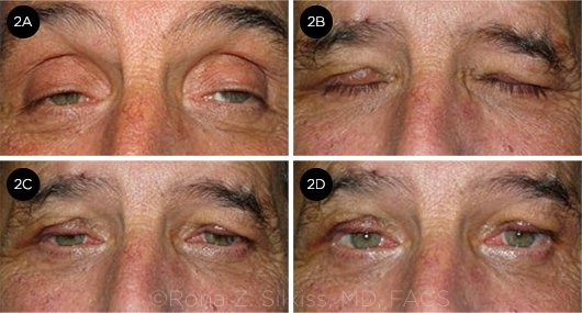
RESULTS. (2A) Pre-op and (2B-2D) one week after silicone rod frontalis suspension.
|
Management
Mueller’s muscle resection, levator resection, and frontalis suspension are the surgical treatments for ptosis in CPEO patients. We chose to treat our patient with a frontalis suspension because of the severity of his ptosis and muscle weakness (Fig. 2).
Frontalis suspension. Frontalis suspension is a frequently used surgical technique to correct moderate to severe drooping of the upper eyelid in the presence of poor or absent levator muscle function (6 mm or less). In this procedure, the connection between the eyelid margin and eyebrow is enhanced using a sling-type material as a suspender, allowing the frontalis muscle to elevate the eyelid more effectively.
Frontalis suspension is commonly used to treat blepharoptosis secondary to CPEO, cranial nerve III palsy, blepharophimosis syndrome, Marcus Gunn jaw-winking, congenital fibrosis syndrome, muscular dystrophy, myasthenia gravis, double elevator palsy, and aponeurotic ptosis in elderly patients. The operation is usually performed under general anesthesia in children, but it can be performed under local anesthesia with sedation in adults.5
Post-op. Patients may not be able to close their eyelids during sleep for a few weeks to several months after surgery. Lubrication may be needed to avoid exposure keratopathy, especially in adults with CPEO, who often have a poor Bell’s phenomenon. Ptosis recurrence rates after a frontalis procedure have been reported to be as low as 5% with the use of autogenous fascia lata and 8% with the use of banked fascia lata.6,7
Complications. Thigh wound hematomas or muscle herniation can occur during harvesting of autogenous fascia lata. The use of nonautogenous silicone rods can infrequently lead to a potential foreign body tissue reaction (such as granuloma formation), extrusion, and wound infection. However, in general, frontalis suspension is an effective technique for severe congenital and acquired ptosis.8
Our patient. The bilateral frontalis suspension was successful. It improved both his head tilt and the diminished visual field caused by the ptosis.
___________________________
* Patient name is fictitious.
___________________________
1 Yu-Wai-Man P et al. Invest Ophthalmol Vis Sci. 2014;55(13):5109.
2 Heighton JN et al. Mitochondrion. 2019;44:15-19.
3 Lee AG, Brazis PW. Curr Neurol Neurosci Rep. 2002;2(5):413-417.
4 Tarnopolsky M. Chronic progressive external ophthalmoplegia (CPEO). In: Saneto R et al., eds. Mitochondrial Case Studies. Elsevier;2016:49-53.
5 Finsterer J. Aesth Plast Surg. 2003;27(3):193-204.
6 Wagner RS et al. Ophthalmology. 1984;91(3):245-248.
7 Crawford JS. J Pediatr Ophthalmol Strabismus. 1982;19(5):253-255.
8 Pacella E et al. PLoS One. 2016;11(9):e0160827.
___________________________
Dr. Moradi is a fourth-year ophthalmology resident at California Pacific Medical Center. Dr. Silkiss is chief of Oculofacial Plastic Surgery at California Pacific Medical Center in San Francisco. Financial disclosures: None.