Blink
Bietti Crystalline Dystrophy
By João Rosendo, MD, Hospital Espírito Santo EPE, Évora, Portugal
Download PDF

A 47-year-old woman was referred to us with complaints of diminished visual acuity and nyctalopia. BCVA was 20/25 in the right eye and 20/20 in the left. Her biomicroscopy was normal. She had no relevant medical history.
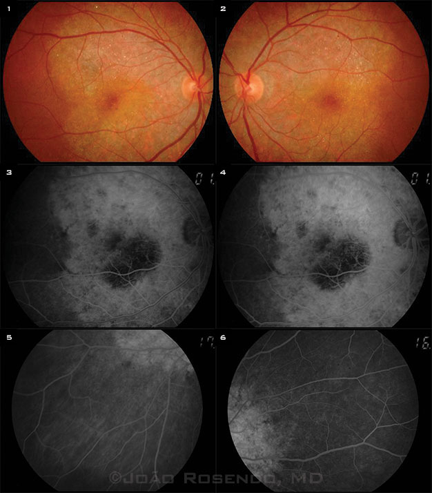
Fundus examinations of each eye showed numerous small white-yellowish crystals in the posterior pole and large confluent areas of retinal pigment epithelial atrophy (Figs. 1 and 2). Fluorescein angiography confirmed posterior pole atrophy of the pigmentary epithelium of the retina and the underlying choriocapillaris (Figs. 3 and 4). Sparing of the peripheral retina was also noticeable (Figs. 5 and 6). There were no important changes in the major retinal vessels or in the optic disc. Static perimetry revealed bilateral central scotomata. Kinetic perimetry and electrophysiologic testing were not possible in this case. The fundus appearance was consistent with Bietti crystalline dystrophy.
| BLINK SUBMISSIONS: Send us your ophthalmic image and its explanation in 150-250 words. E-mail to eyenet@aao.org, fax to 415-561-8575, or mail to EyeNet Magazine, 655 Beach Street, San Francisco, CA 94109. Please note that EyeNet reserves the right to edit Blink submissions. |