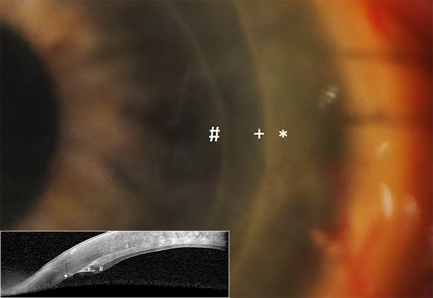
Blink
Can You Guess July's Mystery Condition?
Download PDF
Make your diagnosis in the comments, and look for the answer in next month’s Blink.

Last Month’s Blink
Tiny Bubbles: Perfluorocarbon
Written by Atalie C. Thompson, MD, MPH, and Sharon Fekrat, MD, Duke University Medical Center, Durham, N.C. Photos by Joseph Halabis, OD, Durham Department of Veterans Affairs Medical Center, Durham, N.C.

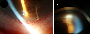
A 53-year-old man presented with an IOP of 29 mm Hg in his right eye eight months after repair of a rhegmatogenous retinal detachment in the same eye. Residual perfluorocarbon (PFC) bubbles with a characteristic “fish egg” appearance were observed in the inferior angle on slit-lamp exam (Fig. 1) and gonioscopy (Fig. 2). On dilated fundus exam, the patient was noted to have developed asymmetric cupping, with a cup-to-disc ratio of 0.6 in the right eye and 0.4 in the left eye. Optical coherence tomography showed mild thinning of the nasal retinal nerve fiber layer in the right eye only. After a discussion with the patient, we removed the PFC bubbles through a paracentesis without complication. His IOP improved to 14 mm Hg after he began dorzolamide-timolol drops twice daily in the right eye.
Retained PFC in the anterior chamber is a rare but serious cause of elevated IOP after retinal surgery. PFC bubbles not only physically obstruct aqueous outflow through the trabecular meshwork but may also cause toxic damage to the delicate structures of the angle. If PFC collects in the anterior chamber, sustained IOP elevation and glaucomatous optic nerve damage may result.
Read your colleagues’ discussion.
| BLINK SUBMISSIONS: Send us your ophthalmic image and its explanation in 150-250 words. E-mail to eyenet@aao.org, fax to 415-561-8575, or mail to EyeNet Magazine, 655 Beach Street, San Francisco, CA 94109. Please note that EyeNet reserves the right to edit Blink submissions. |