Blink
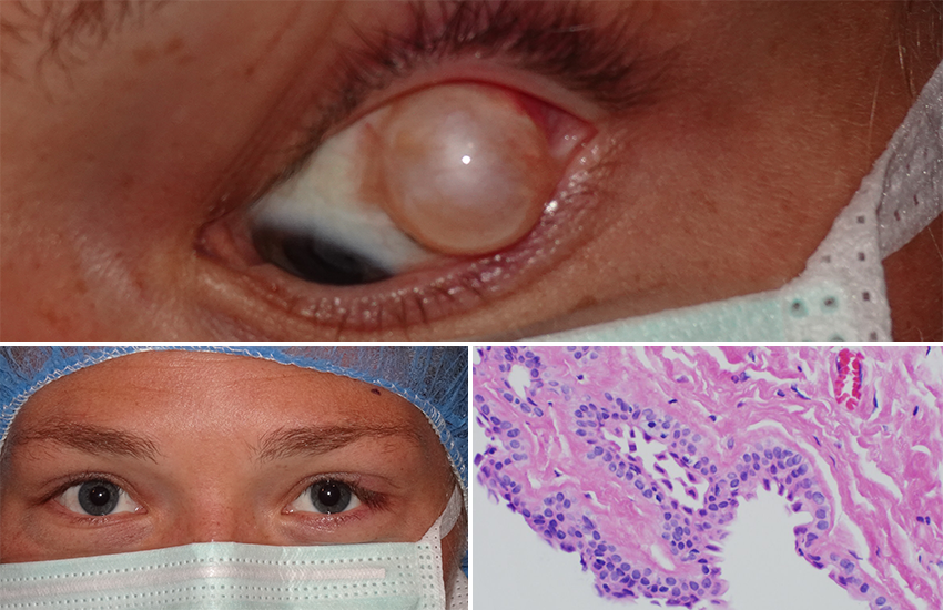
Can You Guess November's Mystery Condition?
Download PDF
Make your diagnosis in the comments, and look for the answer in next month’s Blink.

Last Month’s Blink
Sub-Internal Limiting Membrane Dehemoglobinized Hemorrhage
Written by Danny A. Mammo, MD, and Sandra R. Montezuma, MD, University of Minnesota, Department of Ophthalmology & Visual Neurosciences, Minneapolis. Photo by Drew Miller, University of Minnesota.

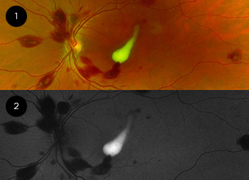
A 32-year-old man with acute myeloid leukemia presented with a one-month history of a large central scotoma of the left eye. His visual acuity was 20/25 in the right eye and count fingers at 2 feet in the left. Posterior segment exam of both eyes demonstrated no anterior vitreous cells, few posterior pigmented vitreous cells, multiple areas of Roth spots, preretinal hemorrhages, and intraretinal hemorrhages. The left fundus also had a white foveal lesion (Fig. 1). On fundus autofluorescence imaging, the lesion was dramatically hyperautofluorescent (Fig. 2). On OCT, it was hyperreflective and was found to be beneath the internal limiting membrane (ILM), consistent with a diagnosis of sub-ILM dehemoglobinized hemorrhage. The patient’s hemorrhages are secondary to his severe leukemia-induced anemia (hemoglobin, 7.2 g/dL) and thrombocytopenia (9). Although hemorrhage is classically hypoautofluorescent on fundus autofluorescence, sub-ILM dehemoglobinized hemorrhage is thought to be hyperautofluorescent because of buildup of fluorophorescent bilirubin due to hemoglobin degradation.
Read your colleagues’ discussion.
| BLINK SUBMISSIONS: Send us your ophthalmic image and its explanation in 150-250 words. E-mail to eyenet@aao.org, fax to 415-561-8575, or mail to EyeNet Magazine, 655 Beach Street, San Francisco, CA 94109. Please note that EyeNet reserves the right to edit Blink submissions. |