Automated Identification of Diabetic Retinopathy Using Deep Learning
By Lynda Seminara and selected by Stephen D. McLeod, MD
Journal Highlights
Ophthalmology, July 2017
Download PDF
As many cases of diabetic retinopathy (DR) are undiagnosed and untreated, improvements in screening and detection are needed. Gargeya and Leng developed technology to automate DR screening and found that a data-driven grading algorithm based on artificial intelligence (AI) and fundus photography can help identify patients with diabetes who require further evaluation and treatment.
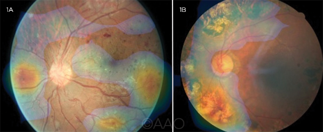
The authors obtained 75,137 publicly available fundus images from patients with diabetes. The images were used to develop and test an AI model capable of differentiating healthy fundi from those with DR. For external validation, the model was tested using images from the Messidor 2 and eOphtha databases. The information gleaned via the automated screening was then displayed through an automatically generated abnormality heatmap, and specific subregions of each fundus image were highlighted for further clinical review.
 |
DEEP LEARNING VISUALIZATION MAPS. Fundus heatmaps overlaid on fundus images, highlighting pathologic regions (1A) in the nasal and temporal quadrants and (1B) in the upper and lower left quadrants.
|
The testing procedure trained 5 separate models, each holding out a distinct validation pool of approximately 15,000 images. Average metrics were derived from 5 test runs on respective held-out data by comparing the model’s predictions with the gold standard determined by the panel of retina specialists. Area under the receiver operating characteristic curve (AUC) was selected as the metric to measure the precision-recall trade-off of the algorithm. Sensitivity and specificity also were calculated.
The model achieved an AUC of 0.97, with 94% sensitivity and 98% specificity on 5-fold cross-validation using the local data set. Testing against the independent Messidor 2 and eOphtha data yielded AUC scores of 0.94 and 0.95, respectively.
The authors concluded that their algorithm is a reliable tool for automated detection of DR. It also offers ease of use, as it can be run on personal computers and smartphones.
The original article can be found here.