This article is from July 2012 and may contain outdated material.
Endophthalmitis is one of the most serious complications following intraocular surgery. In the setting of glaucoma surgery, the risk of endophthalmitis extends well beyond the immediate postoperative period. It is critical that both patients and physicians recognize the signs and symptoms of infection, with the understanding that these may present even years after surgery.
Infections after glaucoma surgery—either trabeculectomy or placement of a glaucoma drainage device (GDD)—can be divided into two groups according to onset: acute infections, which appear within the first four to six weeks, and delayed infections, which occur after six weeks. Post-trabeculectomy infections may be further classified as blebitis (without vitreous involvement) or as endophthalmitis, which includes vitreous involvement and has a worse prognosis.
Reports of the overall incidence of trabeculectomy-associated endophthalmitis vary with surgical technique and use of antimetabolite; one literature review places it in the range of 0.12 to 1.2 percent per year.1 In a retrospective study of Medicare claims data, the cumulative incidence of endophthalmitis in the first year after GDD placement was 0.4 percent.2 The Tube Versus Trabeculectomy (TVT) study, a randomized trial of GDD surgery versus trabeculectomy with mitomycin C as a second surgery for patients with glaucoma, provides the only direct comparison of endophthalmitis rates associated with these surgical interventions. In the TVT, endophthalmitis developed in 1 of 107 eyes in the GDD group and 5 of 105 in the trabeculectomy group over five years.3
Endophthalmitis requires urgent attention, as complete loss of vision can occur quickly. This review will highlight the pathogenesis, presentation, and treatment of endophthalmitis after glaucoma surgery.
Microbiology
The organisms responsible for endophthalmitis vary according to source. Endogenous infections develop from distant sites of infection within the patient’s body, such as the urinary tract or heart valves. Exogenous infections result from the introduction of microbes into the eye via surgery or trauma. In the case of cataract surgery, the organisms most commonly seen are those that often colonize the ocular surface: coagulase-negative staphylococci (70 percent), followed by Staphylococcus aureus (10 percent) and streptococci (9 percent).4
In post-trabeculectomy endophthalmitis, however, the most common pathogens are streptococci in acute endophthalmitis and both streptococci and Haemophilus influenzae in delayed endophthalmitis.4 In addition to H. influenzae, gram-negative organisms have also been isolated in cases of post-trabeculectomy endophthalmitis.4 Infections resulting from gram-negative species and streptococci are associated with worse visual outcomes (only 45 or 46 percent of eyes measured 20/400 or better) than those resulting from coagulase-negative staphylococci (89 percent of eyes measured 20/400 or better).5
Organisms (commonly staphylococci) associated with post-GDD endophthalmitis are similar to those seen in endophthalmitis following cataract surgery, perhaps due to the seeding of bacteria from the ocular surface, exacerbated by conjunctival erosions over the tube or plate of the GDD.6 Rarely, fungus has been implicated as the causative pathogen.
 |
|
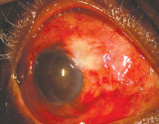
Complication. A patient with bleb-associated endophthalmitis.
|
Risk Factors
The use of antimetabolites, including mitomycin C and 5-fluorouracil, reduce the risk of bleb failure due to scarring but may increase the risk of postoperative infection.4 Because the potential benefits outweigh the risk, adjunctive use of antimetabolites with trabeculectomy surgery is common practice. Note that the location of the filtering bleb affects the risk of infection: Placement of a bleb inferior to the horizontal meridian should be avoided, because it is more likely to lead to endophthalmitis than is one located superiorly.4
A history of bleb leak is common in eyes with post-trabeculectomy endophthalmitis. Although the management of bleb leaks varies greatly by surgeon, a patient with a recurrent or persistent leak is at greater risk for infection and should be made aware of this possibility.
In GDD surgery, no correlation has been found between the type of device and the incidence of infection. However, multiple studies have shown a higher rate of endophthalmitis in patients younger than 18 years of age.6 Erosion of the conjunctiva over the tube is a risk factor, as is reoperation, including repositioning of the tube, capsulectomy, and needling.6 Inspection of the conjunctiva over the tube is recommended at every visit, as areas of conjunctival dehiscence can be small and easily missed. As with bleb leaks, the management of tube exposure varies with the clinical scenario, but any suspected erosion should be evaluated by a glaucoma surgeon, as operative repair is generally required.
Signs and Symptoms
Glaucoma patients with postsurgical endophthalmitis may present with pain or decreased vision, but the onset of symptoms may be more insidious than that generally seen in patients with endophthalmitis following cataract surgery.5 Initial symptoms can be similar to uveitis, including mild pain and photophobia.4 In a case series of trabeculectomy-associated endophthalmitis, 35 percent of subjects demonstrated prodromal symptoms such as brow ache, headache, or inflammation of the ocular adnexa in clinic visits before diagnosis.1
On examination, the patient is likely to demonstrate decreased visual acuity, along with inflammation involving the conjunctiva, anterior chamber (often with hypopyon), and vitreous. In endophthalmitis following trabeculectomy, accumulation of cellular debris and pus may cause the conjunctival bleb to appear opaque; in the setting of otherwise diffuse conjunctival injection, this gives rise to the classic “white on red” appearance.1 A bleb leak may or may not be present. Leaks may sometimes be masked by debris plugging the conjunctival defect or by hypotony, which makes the leak intermittent. In the case of an opacified anterior segment, ultrasound helps detect vitreous inflammation and diagnose retinal detachment, and it provides a basis for subsequent treatment.4
Management
Sampling of the intraocular fluid for Gram stain and culture is recommended before treatment is initiated. However, empiric treatment should begin promptly while the specimen is being analyzed.
Obtaining a specimen. It is not uncommon to have negative culture results even when the clinical diagnosis of infectious endophthalmitis is obvious. Vitreous tap provides the best method for obtaining a positive culture specimen and is often combined with an injection for treatment. Collecting superficial pus from the conjunctiva is more likely to yield mixed flora and should not substitute for acquiring intraocular fluid for analysis.
Antibiotics. The initial choice of antibiotics for bleb-associated endophthalmitis should cover a broad spectrum. Local availability of medication and local resistance patterns will guide the selection. A common combination for intravitreal treatment is vancomycin, which provides gram-positive coverage, and a third-generation cephalosporin such as ceftazidime, for gram-negative activity. Intravitreal amphotericin B, voriconazole, and caspofungin act against fungi.4 The use of intravitreal corticosteroids is controversial; they may reduce early inflammation, but their effect on visual outcome is unclear. Subconjunctival dexamethasone is a reasonable alternative to intravitreal corticosteroids.4
In addition to intravitreal therapy, we recommend broad-spectrum topical antibiotic drops until culture results are available to guide more specific therapy. Vancomycin 25 mg/mL and amikacin 20 mg/mL, alternating on the hour, is a common regimen. The addition of a topical corticosteroid may help reduce inflammation.
Vitrectomy. The role of vitrectomy in the setting of endophthalmitis after glaucoma surgery is a matter of debate. Although the Endophthalmitis Vitrectomy Study (EVS) provides guidelines with respect to outcomes following endophthalmitis treatment with intravitreal injection versus initial pars plana vitrectomy, the population studied in the EVS did not include patients with post–glaucoma surgery endophthalmitis; and extrapolation of its recommendations to this population is not appropriate.6 Given the more virulent organisms found in bleb-associated endophthalmitis, it may be reasonable to consider vitrectomy earlier in these patients than in patients with endophthalmitis following cataract surgery, but evidence-based data to drive this clinical decision are lacking.
Removing a GDD. Removal of an implanted GDD is also controversial. Some investigators have suggested removing the device because it may serve as a reservoir for the offending organism. Others cite successful outcomes without removal of the device.6
Ex-Press mini-shunt. A relatively recent development in glaucoma surgery is the Ex-Press mini-shunt (Alcon), which is placed under the conjunctival flap in filtering surgery. There is little specific evidence to direct the management of endophthalmitis in the setting of an Ex-Press mini-shunt. Whether or not the Ex-Press shunt is explanted from an eye with endophthalmitis, the drug strategy should be similar to other scenarios: broad-spectrum antibiotic coverage, with more specific tailoring of treatment as culture results become available.
Summary
Appropriate vigilance facilitates the prompt recognition and treatment of endophthalmitis following glaucoma surgery. Clinicians should search diligently for bleb leaks and conjunctival erosions in patients who have undergone glaucoma surgery. Patients should be instructed to seek care immediately if ocular redness, pain, discharge, or decreased vision develops. If the clinical exam supports the diagnosis of endophthalmitis, sampling of intraocular fluid for culture followed by administration of broad-spectrum antibiotics is indicated. Ongoing research should help clarify the role of vitrectomy and removal of implanted drainage devices in the future.
___________________________
1 Ang GS et al. Br J Ophthalmol. 2010;94(12):1571-1576.
2 Stein JD et al. Ophthalmology. 2008;115(7):1109-1116.
3 Gedde S et al. Am J Ophthalmol. 2012;153(5):804-814.
4 Lemley CA, Han DP. Retina. 2007;27(6):662-680.
5 Jacobs DJ et al. Clin Ophthalmol. 2011;5:739-744.
6 Wentzloff JN et al. Int Ophthalmol Clin. 2007;47(2):109-115.
___________________________
Mr. Farber is a fourth-year medical student at the University of Virginia, and Dr. Muir is an assistant professor of ophthalmology at Duke University and the Durham VA Medical Center. The authors report no related financial interests.