Download PDF
Manual devices and surgical microscopes that can perform optical coherence tomography (OCT) during vitreoretinal surgery have been available in the United States for a few years. Now, a new fiberoptic probe, made for microinsertion into the eye, can expand the capabilities of intraoperative OCT, Japanese researchers have found.
“Handheld-type and scope-mounted OCT give us images of intraoperative retinal configurations, mainly in the posterior retina. On the other hand, this intraocular fiber-type OCT enables us to obtain images of tissues anywhere in the eye,” said Hiroko Terasaki, MD, PhD, professor and chairman of ophthalmology at the Nagoya University, in Nagoya, Japan. Dr. Terasaki and coauthors reported last summer on their early clinical testing of the fiberoptic system.1
Probe fits through microincision. The fiberoptic, swept-source OCT probe (Nidek) is inserted through the same type of 23-gauge trocar used for microincision vitrectomy. Its scanning laser beam is at a 43-degree angle to the visual axis. By rotating the fiberoptic, the surgeon can visualize not only the posterior retina but also the peripheral retina near the trocar, the ciliary body, and tissues in the anterior chamber.
The images are displayed in real time on a screen mounted next to the ocular of the operating microscope. The device has axial and lateral resolutions of 3.65 μm and 80 μm, respectively, and the scanning laser’s central wavelength is 1,060 nm. During pilot clinical testing in 3 patients, the researchers said, this wavelength enabled them to acquire detailed information on deeper retinal and choroidal tissues, including the optic disc.
 |
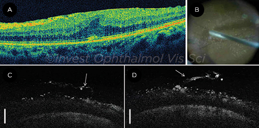
PATIENT WITH ERM. (A) Desktop OCT shows an ERM and macular edema. (B) Intraoperative microscope image shows OCT probe in place under chandelier illumination. (C, D) OCT images of a half-peeled ERM and the internal limiting membrane. Particles of triamcinolone are seen on the retina and the ERM. Scale bars: 500 μm.
|
Can aid in surgical decision making. “The image quality may not be sufficient for a precise analysis of retinal thickness. However, it should give surgeons adequate information to make a decision on how to proceed with intraoperative procedures,” they wrote.
For example, the group presented the case of a patient with epiretinal membrane (ERM). The fiberoptic OCT probe was able to show the half-peeled ERM, particles of triamcinolone acetonide, and the incision site with the trocar still in place (see Figure).
Limitations. However, the researchers noted several current limitations:
- The surgeon must take care to prevent intraocular injury from the probe tip, by holding it 1.5 mm to 2 mm away from the target tissue.
- The surgeon’s eyes must move about 30 degrees to the side to view the OCT display screen, which could cause a loss of the microscope view.
- The images lose contrast if illumination in the optical system decreases or if there are motion artifacts from rotation of the fiberoptic inside its cable.
These limitations may be overcome in the near future, said Dr. Terasaki, by development of a new 25-gauge fiberoptic probe, heads-up surgery, and improvement of the OCT system.
Applications. According to Dr. Terasaki, “Intraoperative OCT allows the surgeon to detect a tissue abnormality that was obscured by a media opacity before surgery or to see how the surgery itself changed the retinal condition. For instance, intraoperative OCT can reveal that there was a retinal tear during surgery, which may prevent the need for a second procedure later.”
—Linda Roach
___________________________
1 Asami T et al. Invest Ophthalmol Vis Sci [Special Issue]. 2016;57(9):OCT568-574.
___________________________
Relevant financial disclosures—Dr. Terasaki: Carl Zeiss Meditec: L; Nidek: L, P, S.
For full disclosures and disclosure key, see below.

More from this month’s News in Review