Download PDF
This month, News in Review Highlights selected papers from the original papers sessions at AAO 2017. Each was chosen by the session chair because it presents important news or illustrates a trend in the field. Only 4 subspecialties are included here; papers sessions will also be held in 6 other fields. See the Meeting Program, which you’ll find in your meeting bag, or the Mobile Meeting Guide for more information.
The lamina cribrosa is regarded as the primary site of pathogenesis in glaucoma. Now there’s evidence that the lamina cribrosa (LC) may also be the site of an early warning system for subsequent visual field (VF) loss.
Study rationale. LC deformation has been noted to occur primarily in early glaucoma stages, and biomechanical changes to the LC appear to be closely associated with glaucomatous change. “This study was undertaken to investigate the association between the extent of baseline LC deformation and subsequent glaucoma progression,” said Ahnul Ha, MD, who is at Seoul National University Hospital in South Korea.
The researchers evaluated 101 eyes with early-stage primary open-angle glaucoma (POAG). All eyes had been followed longer than 3.5 years, had undergone more than 5 reliable standard automated perimetry tests, and had well-controlled intraocular pressure (IOP) during follow-up.
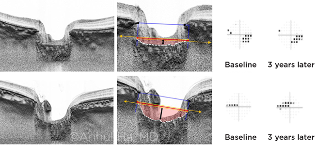
Using swept-source optical coherence tomography (SS-OCT), the researchers took baseline LC images and then calculated manually what they call the lamina cribrosa curvature index (LCCI) at 12 radial lines.
 |
PROGRESSION. The researchers used SS-OCT to calculate the LCCI (lamina cribrosa curvature index) at baseline and compared that to later visual field changes.
|
Faster progression. Baseline LCCI showed a significant correlation with the rate of subsequent VF progression. Eyes with greater LCCI values (eyes with greater LC deformation) at early stages of glaucoma showed a significantly faster rate of VF progression, even in cases with well-controlled IOP.
Higher risk in younger patients. A separate analysis by age group revealed significantly faster VF progression, with LCCI changes occurring more rapidly in the relatively younger age group (< 68 years). Dr. Ha speculated this could be due to the more pliant LC microstructure of younger individuals, which may cause “more kinking and pinching of the axons in the laminar pores,” inducing faster progression.
| Baseline Lamina Cribrosa Curvature Index and Prediction of Glaucoma Progression. When: Monday, Nov. 13, 2:48-2:55 p.m., during the glaucoma original papers session (2:00-5:00 p.m.). Where: Room 271. Access: Free. |
Clinical implications. The findings suggest that axons of retinal ganglion cells might be more vulnerable to further glaucomatous injury in POAG eyes with greater posterior bowing of the LC. And while the results suggest that greater LCCI value can be regarded as a risk factor for further progression in POAG, Dr. Ha stressed that the LCCI value is only a single parameter of the LC’s deformation and is not a direct index of glaucoma progression.
The bottom line, she said: “In vivo assessment of the LC by SS-OCT is becoming more important, so that these eyes can be monitored more carefully for subsequent VF deterioration.”
—Miriam Karmel
___________________________
Relevant financial disclosures—Dr. Ha: None.
For full disclosures and disclosure key, see below.

More from this month’s News in Review