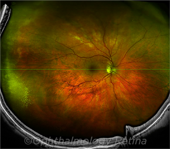
Nomenclature and Guidelines for Widefield Imaging
By Jean Shaw
Selected By: Andrew P. Schachat, MD
Journal Highlights
Ophthalmology Retina, October 2019
Download PDF
The term widefield has been used inconsistently in the literature in describing retinal images. To address this problem, an international panel of 11 retina specialists was convened to evaluate the terminology for widefield images obtained with current retinal imaging methods. Choudhry et al. summarized the findings and recommendations of the panel, known as the International Widefield Imaging Study Group.
Before the consensus meeting, a set of seven images was circulated to panel members. The images were acquired with current approved and commonly used imaging methods, including swept-source optical coherence tomography (OCT) and fluorescein angiography. Both healthy and diseased eyes were represented in the image set.
 |
DEFINITIONS. The panel proposed that the term panretinal be used for a complete 360-degree ora-to-ora view of the retina. In lieu of a single capture method, the montage technique (shown here) is likely to remain popular.
|
In addition to evaluating the capabilities of the imaging methods, panel members also reviewed published studies and discussed the definitions of widefield and ultra-widefield imaging. Their recommendations include the following:
- The term widefield should be limited to images depicting retinal anatomic features beyond the posterior pole, but posterior to the vortex vein ampulla, in all four quadrants.
- Ultra-widefield images are those showing retinal anatomic features anterior to the vortex vein ampulla in all four quadrants.
- Posterior pole should continue to be defined as the area of retina within the major temporal vascular arcades and slightly just beyond, as captured in a 50-degree view with most devices.
- In discussing field of view (FOV) and its point of centration for fundus cameras and OCT devices, the panel agreed that the FOV should have the macula at its center.
- FOV boundaries should correspond to anatomic landmarks within the retina, and these landmarks should be incorporated into the nomenclature. For instance, they said, the term midperiphery should refer to the region of retina extending from the vascular arcades to the posterior edge of the vortex vein ampulla, which is captured at a range of 60 degrees to 100 degrees.
Finally, panel members proposed that this standardized nomenclature be used in future publications, and they noted that their findings will need to be updated in the future as imaging technology develops.
The original article can be found here.