Ocular Side Effects of MEK Inhibitors: Fluid Foci
By Lynda Seminara
Selected By: Stephen D. McLeod, MD
Journal Highlights
Ophthalmology, December 2017
Download PDF
Francis et al. studied the characteristics of serous retinal disturbances in patients who receive the class of drugs known as mitogen-activated protein kinase (MEK) inhibitors. They found that certain features distinguish these disturbances from those noted in central serous chorioretinopathy (CSC), even though the conditions have been considered analogous by some researchers.
For this retrospective single-center study, the researchers included 313 fluid foci from a total of 25 patients (50 eyes) who were receiving MEK inhibitors to treat metastatic cancer. All eyes had evidence of serous retinal detachment, confirmed by optical coherence tomography (OCT). The researchers assessed the presence or absence of subretinal fluid via clinical examination and OCT, and they evaluated the morphology, distribution, and location of fluid foci serially for each eye.
Two independent observers measured choroidal thickness at 3 time points (baseline, fluid accumulation, and fluid resolution). Statistical analysis was used to correlate interobserver findings and to compare choroidal thickness and visual acuity at each time point. OCT characteristics of retinal anomalies at baseline were compared with those at fluid accumulation.
 |
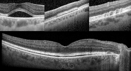
FLUID FOCI CONFIGURATIONS. Domes (upper left) appear as dome-shaped fluid accumulation between the RPE and the interdigitation zone. Caterpillar foci (upper middle) appear as straight or plateaued low-lying accumulations. Wavy foci (upper right) present as a linear collection of tiny domes and displace the interdigitation zone in a wave-like pattern. Splitting foci (lower) appear as a broad, low-lying accumulation of fluid between the RPE and the interdigitation zone.
|
Most patients (92%) had bilateral fluid foci, which is less common in CSC. Most fluid foci in this study (77%) were multifocal, with at least 1 focus involving the fovea (83%). All fluid foci occurred between the interdigitation zone and an intact retinal pigment epithelium (RPE). Regarding morphology, the 313 fluid foci were classified as follows: dome (n = 231; 73.8%), caterpillar (n = 36; 11.5%), wavy (n = 31; 9.9%), and splitting (n = 15; 4.8%). Best-corrected visual acuity at fluid resolution did not differ significantly from that at baseline, and no eye lost more than 2 Snellen lines from baseline to fluid accumulation.
Choroidal thickness was similar at the 3 time points. Interobserver correlations were strong for choroidal thickness measurements and morphology grading. Contrary to typical CSC findings, the retinal pigment epithelium and choroid remained normal during MEK inhibition. There was no irreversible loss of vision and no serious eye damage.
The authors concluded that the subretinal fluid foci caused by MEK inhibition appear clinically and morphologically unique, and they noted that large prospective studies with greater imaging frequency are needed to draw firm conclusions.
The original article can be found here.