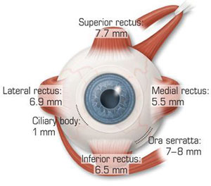
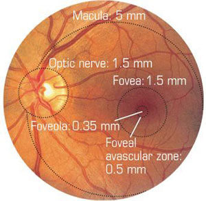
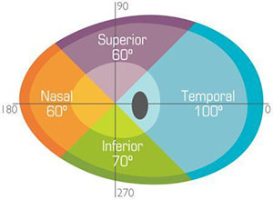
the most basic ophthalmic exam to a complicated cataract surgeries, expertise in eye anatomy is the building block of everything that is ophthalmology. Here are a few of the more elemental ophthalmic measurements that all residents should put to memory — from orbital volume and posterior segment diameters to optic nerve length and visual fields.
Orbital entrance: 35 mm W x 40 mm H
Orbital volume: 30 mL
Globe volume: 6.5 mL
Anterior chamber volume: 250 µL
Conjunctival sac volume: 35 µL
Aqueous humor forms: 2–2.5 µL/min
Aqueous turnover rate: 1%/min
Basal tear secretion: 2 µL/min
Eyedrop volume: 50 µL
Distance From the Limbus

Corneal thickness: 500–600 µm
K horizontal diameter (infant): 10 mm
K horizontal diameter (adult): 11.5 mm
Lens diameter: 9.5 mm
Anterior capsule thickness 14 µm
Posterior capsule thickness 2–4 µm
Ciliary sulcus diameter: 11.0 mm
Posterior Segment

Rod photoreceptor cells: 120 million
Cone photoreceptor cells: 6 million
Retinal ganglion cells (at birth): 1.2 million
52% of optic nerve fibers cross in optic chiasm (nasal fibers)
Optic Nerve Length
Monocular Visual Field

* * *
About the author: James G. Chelnis, MD, is preparing for his oculoplastics fellowship, which starts in July at Vanderbilt University. He is currently a senior resident at the University at Buffalo, where he graduated from medical school and was the school government president. In this role, he helped renegotiate the university’s health insurance terms with their provider, create a nationwide student–alumnus network, and organize a “Career Day” to place students in direct contact with physicians of all specialties prior to clinical rotations.