Download PDF
Make your diagnosis in the comments, and look for the answer in next month’s Blink.

Last Month’s Blink
Traumatic Aniridia
Written by Alanna S. Nattis, DO, Lindenhurst Eye Physicians and Surgeons, Babylon, N.Y.; Henry D. Perry, MD, Nassau University Medical Center, East Meadow, N.Y.; Eric D. Donnenfeld, MD, New York University, New York, N.Y.; Eric D. Rosenberg, DO, New York Medical College, Valhalla, N.Y. Photo by Henry D. Perry, MD.

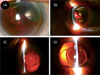
A 77-year-old woman, 2 weeks after undergoing uncomplicated laser-assisted phacoemulsification of her right eye, presented 2 hours after falling onto the right side of her face. Her vision was hand motion with 2+ corneal folds, total hyphema, and IOP of 40 mm Hg. There was no evidence of corneal or scleral laceration. Upon emergent anterior chamber washout (1A; note residual blood), absence of the iris was observed (1B). No vitreous hemorrhage or retinal detachment was evident. The recently implanted IOL was centered and intact within the capsular bag. The patient was placed on topical glaucoma medications, bed rest, and a short course of oral prednisone. Over 1 month, her corneal edemaand hyphema cleared (1C). A year later, with total aniridia and a centered, intact IOL (1D), she denies complaints of glare and has maintained 20/30 uncorrected VA.
There are few reports of isolated aniridia in pseudophakic eyes after nonperforating blunt trauma, without dehiscence/extension of the cataract incision.1-6 Small self-sealing modern cataract incisions appear to be protective against expulsion of intraocular contents, which has led to several theories regarding the mechanism of traumatic aniridia.2-6 Acute rise in IOP from blunt trauma may allow the corneal incision to act as a “release valve,” promoting rapid progression of an iridodialysis to complete avulsion and subsequent expulsion due to the high-velocity injury.3,4 In our case, there was total iris loss, but the remainder of the intraocular contents remained stable and intact. Thus, it is possible that the IOL may have acted to absorb the impact and block disruption of surrounding tissue.5 An additional theory is that the traumatized iris may have remained within the eye, only to undergo rapid phagocytosis by macrophages and trabecular meshwork cells.3
___________________________
1 Gencer B et al. Turk J Ophthalmol. 2014;44:80-82.
2 Kim KH, Kim WS. Arq Bras Oftalmol. 2016;79(1):44-45.
3 Parmeggiani F et al. J Ultrasound Med. 2007;26:1795-1797.
4 Muzaffar W, O’Duffy D. J Cataract Refract Surg. 2006;32(2):361-362.
5 Khemka S et al. The Internet Journal of Ophthalmology and Visual Science. 2005;4(1):1-4.
6 Mikhail M et al. Clin Ophthalmol. 2012;6:237-241.
Read your colleagues’ discussion.
| BLINK SUBMISSIONS: Send us your ophthalmic image and its explanation in 150-250 words. E-mail to eyenet@aao.org, fax to 415-561-8575, or mail to EyeNet Magazine, 655 Beach Street, San Francisco, CA 94109. Please note that EyeNet reserves the right to edit Blink submissions. |