Blink
Posterior Polymorphous Corneal Dystrophy
By Rony R. Sayegh, MD, University Hospitals Case Medical Center, Cleveland
Photo by Mark Harrod, CRA, OCT-C, University Hospitals Case Medical Center, Cleveland
Download PDF

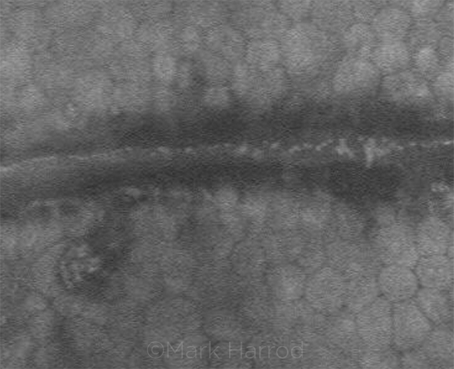
A 60-year-old man was referred to our clinic after a corneal lesion was noted in both eyes during routine examination. Confocal microscopy was obtained (see image).
Posterior polymorphous dystrophy (PPMD) is a rare corneal dystrophy with an autosomal dominant inheritance and great variability in clinical expression. It is usually asymptomatic, although corneal edema can occasionally be present. Peripheral anterior synechiae, iris abnormalities, corectopia, and elevated intraocular pressure may also occur. PPMD can appear in 3 possible patterns: vesicle-like lesions, band lesions (as in our patient), and diffuse opacities. Polymegathism and some pleomorphism of the endothelial cells lining the band are visible on confocal microscopy. The band consists of shallow trenches and ridges arising from a large number of confluent vesicles. Bright, prominent, well-defined endothelial nuclei can sometimes be seen. Confocal microscopy can be helpful in differentiating a very asymmetric presentation of PPMD from iridocorneal endothelial syndrome. Corneal transplantation can be performed for endothelial decompensation.
| BLINK SUBMISSIONS: Send us your ophthalmic image and its explanation in 150-250 words. E-mail to eyenet@aao.org, fax to 415-561-8575, or mail to EyeNet Magazine, 655 Beach Street, San Francisco, CA 94109. Please note that EyeNet reserves the right to edit Blink submissions. |