We all love ophthalmology because we are able to directly visualize the pathology and watch its resolution through our expert management — and the slit lamp is an essential tool.
Keep in mind that slit lamps are all a little different, so familiarize yourself with them at each clinic’s location. Don’t be afraid to ask for help on day 1!
Here are some expert tips on what to look for to ensure you are using the instrument correctly.
Ergonomics
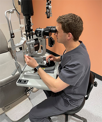
Pay attention to positioning, both for yourself and your patient. Adjust the height of the slit lamp so you are sitting with a straight back. Then adjust the chin rest so your patient’s eyes line up with the black line on the chin rest holder.
Instruct the patient to keep their forehead pressed
against the head rest. Place your dominant hand on the joystick and your nondominant hand on the illumination system arm. This will allow you to have excellent control of your slit lamp.
For challenging patients with a large chest or abdomen: These body parts can get in the way of the slit lamp. So lift their chair up high and have them come forward in the seat and lean forward, pivoting at the waist.
 |
|
Dr. Silverstein models the correct posture and hand positions at the slit lamp.
|
Be Systematic
Use the same system for every patient, every time. If you don’t follow this rule, you’ll miss important examination findings.
Start with the right eye: Eyelids, eyelashes, meibomian glands, palpebral conjunctiva, upper lid and then lower lid.
Conjunctiva and sclera: Have the patient look in all directions so you can examine all parts of the anterior globe.
Cornea: Start with diffuse light, and then bring the light directly in front of the eye for a red reflex. This will highlight irregularities in the cornea as well as the lens and will show transillumination defects in the iris.
Now, narrow your beam (with your nondominant hand already resting on the width control). Examine the epithelium, then the stroma and then the endothelium. Keep your light temporal for the temporal side of the cornea and swing your light nasally for the nasal examination.
Pro tip: Lift the upper lid to examine the superior cornea. Here PGY-2s often miss significant pathology.
Anterior chamber (AC): Look for cell/flare by focusing on the iris, moving over the pupil and then pulling toward you slightly. Reduce the size of your beam to 1 by 1 mm. Use increased magnification. Also make sure the AC is deep — if shallow, don’t apply dilating drops.
Iris: Look for neovascularization and nevi.
Lens: Repeat the red reflex if necessary. Look at the anterior lens, posterior lens and nucleus.
Anterior vitreous: In patients with floaters, look for pigment in the vitreous, which is a sign of retinal detachments/tears (Schafer’s sign).
Move to the left eye.
Pro tip: Mind the nose! Pull the slit lamp in an arc around the patient.
 |
|
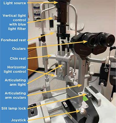
Must-know parts of the slit lamp.
|
Tools of the Lamp
Slit lamps have various components that you will need to familiarize yourself with:
The joystick: Think of this as the “fine focus” of your microscope. Always keep a hand here.
Cobalt blue filter: Learn to switch from white light to blue light. This is different on each slit lamp. Apply your fluorescein after looking at the cornea; the staining can block your view of important corneal findings.
Vertical slit beam: This can be used to measure structures and can rotate in different directions. For example, measure a hyphema or a corneal ulcer.
Rules of the Lamp
Be aware of your lamp’s settings and make sure you always follow these habits.
Reset the ocular optics to zero which can be found on the top of the ocular piece. You can dial in your prescription here if you want to use the slit lamp without glasses.
Turn off the slit lamp and lock it after each use.
Marvel in the beauty of the eye!
Further Resources
Tim Root from Ophthobook.com has a fantastic summary about using the slit lamp on YouTube. youtube.com/watch?v=w9wMJ6job_0.
Slit Lamp Examination. EyeWiki. eyewiki.aao.org/Slit_Lamp_Examination.
How to Use a Slit Lamp. American Academy of Ophthalmology. aao.org/young-ophthalmologists/yo-info/article/how-to-use-slit-lamp.
* * *
 Evan Silverstein, MD,
 is a pediatric ophthalmologist and an assistant professor of ophthalmology and associate resident program director at Virginia Commonwealth University in Richmond, VA.