Download PDF
British researchers have identified unique retinal scarring in survivors of Ebola virus disease (EVD), the distribution of which suggests a neurotrophic spread of the disease into the eye.1
A surprising find. Paul J. Steptoe, BSci, MBChB, and his team conducted a case control study in Freetown, Sierra Leone, to investigate ocular signs in EVD survivors. A total of 82 patients with ocular symptoms and 105 controls underwent ophthalmic examination, including widefield retinal imaging.
 |
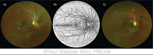
LESIONS. Retinal images (1A, 1C) show Ebola peripapillary and peripheral lesions following the anatomic distribution of the ganglion cell axons (1B). Asterisks in the fundus photos indicate curvilinear lesions distinct from the retinal vasculature. White arrowhead indicates retinal nerve fiber wedge defect.
|
Lesion appearance. “Despite a high prevalence of background retinal disease in our study population, we discovered a novel retinal lesion in almost 15% of survivors,” said Dr. Steptoe, at the University of Liverpool and Royal Liverpool Hospital in Liverpool, U.K. “And although the shape of the lesions varied, we were surprised to find a very unusual angulated appearance, often resembling a diamond or wedge shape.”
Lesion location. These lesions were located either in the fundus periphery or adjacent to the optic disc. When they were near the optic disc, their curvilinear projections from the disc margin aligned with the retinal ganglion cell axons that make up the optic nerve. “This distribution suggests a neuronal transmission of the disease to the retina in a similar fashion to West Nile virus retinitis,” said Dr. Steptoe.
Is cataract surgery safe? Cataract surgery is currently denied EVD survivors for fear of viral persistence in the ocular fluid. To assess the validity of this claim, the researchers also tested the aqueous fluid in 2 survivors who had cataracts but no anterior chamber inflammation; both were negative for the Ebola virus.
“Although larger investigations are ongoing, our study provides provisional evidence that cataract surgery is indeed safe for these EVD survivors,” said Dr. Steptoe. His team is currently conducting a yearlong follow-up to assess the recurrence of EVD as well as any further retinal changes in survivors.
—Mike Mott
___________________________
1 Steptoe PJ et al. Emerg Infect Dis. 2017;23(7):1102-1109.
___________________________
Relevant financial disclosures—Dr. Steptoe: Bayer Global Ophthalmology Awards Programme: S; Optos: S; The Dowager Countess Eleanor Peel Trust: S.
For full disclosures and disclosure key, see below.

More from this month’s News in Review