NOV 20, 2017
A Compendium of Inherited Disorders and the Eye, Oxford University Press
Genetics
OMIM Number
Inheritance
- 95% of cases are sporadic trisomy 211
- 3-4% have unbalanced translocation between chromosome 21 and another chromosome, most often chromosome 14. Approximately three quarters of these are de novo, with the remainder resulting from familial translocations.1
- 1-2% have cellular mosaicism with one normal cell line and one with trisomy 21.1
Gene/Gene Map
- 21q22.32
- 5 Mb between the loci D21S58 and D21S42 have been found to be associated with the mental retardation and facial features of the syndrome. The subregion D21S55, in particular, has been identified as associated with mental retardation, oblique eye fissures, Brushfield spots, epicanthus, flat nasal bridge, protruding tongue, short broad hands, clinodactyly of the fifth finger, a gap between the first and second toes, hypotonia, short stature, and the characteristic dermatoglyphics.2
Epidemiology
- Occurrence increases with maternal age. Incidence at a maternal age of 12-29 years is 1 in 1500 live births, at age 30-34 years it is 1 in 800 live births, at age 35-39 years it is 1 in 270 live births, at age 40-44 years it is 1 in 100 live births, and at older than 45 years, it is 1 in 50 live births.3 Although incidence increases with age, 50-80% of babies with DS are born to mothers younger than 35 years of age, since younger women tend to have more babies in general.4
Clinical Findings
- Down syndrome is associated with multiple malformations and cognitive impairment.
- The most common physical manifestations include hypotonia, a small brachycephalic head, a flat nasal bridge, epicanthal folds, up-slanting palpebral fissures, Brushfield spots, a small mouth, small ears, excessive skin at nape of neck, transverse palmar crease, a short fifth finger with clinodactyly, and a deep plantar groove between the first and second toes.1
- Cognitive impairment can be mild, moderate, or severe.
- 50% have congenital heart defects. The primary lesions identified include complete atrioventricular septal defect, ventricular septal defect, atrial septal defect, partial atrioventricular septal defect, tetralogy of fallot, and patent ductus arteriosus.1
- Approximately 75% have hearing loss, and 70% experience chronic otitis media.1,5
- 50-79% experience obstructive sleep apnea, including those who are not obese.1
- 6-12% have gastrointestinal tract abnormalities including duodenal atresia or stenosis, imperforate anus, and esophageal atresia with tracheoesophageal fistula.6
- 4-18% have thyroid disease.1
- There is an increased risk of type 1 diabetes.5
- Most adults develop dementia and functional changes typical of Alzheimer disease by the sixth decade of life.7
- Less common manifestations include transient myeloproliferative disease (4%-18%), leukemia (1%), Hirschsprung disease (<1%).1
Ocular Findings
1. Refractive error
- The most common underlying causes of refractive error in Down syndrome patients include hypermetropia, myopia, and astigmatism.
- Significant refractive error is found in approximately 50%-80% of children with Down syndrome. The frequency of reported refractive errors varies based on the definition of significant error. Doyle et al found that almost 80% of children with Down syndrome have a spherical equivalent outside the range of -0.50 to +1.00 D.8,9
- Hypermetropia is found in 55-80% of individuals with Down syndrome.8,10
- Myopia is found in 18-59%.8,11
- The prevalence of astigmatism is typically reported as 20-30%.2
- Oblique astigmatism has been found to be the most prevalent type of astigmatism in these patients. In addition, astigmatism has been found to increase with age in patients with Down syndrome.8 A right eye-left eye specificity of the cylinder axis is present likely related to biomechanical factors induced by the upward slanting of the palpebral fissures and intorsion of the cylindrical axis.12 This is supported by studies by Garcia and colleagues where oblique astigmatism and obliquity of the palpebral fissure are correlated.13
- Corneal astigmatism in individuals with Down syndrome has been shown to be predictive of overall refractive astigmatism, as it is in the general population.14
- There is a greater magnitude and wider orientation variation of astigmatism in these individuals when compared to controls, which is most likely due to previously reported differences in corneal and optical components of the eye compared to the general population.14
- Marsack et al showed by comparing autorefraction readings that there is a greater uncertainty in objective measurements of refraction in Down syndrome patients.15
- At birth, there is no significant difference in refractive error between patients with Down syndrome and controls. However, refractive error increases with age in patients with Down syndrome, while it decreases with age in control children. A theory for this difference with age is a failure of emmetropization in individuals with Down syndrome. The reasons for this failure include poor accommodation, decreased time spent doing near work with an increased amount of time outdoors, and changes in the visual cortex.8
2. Accommodation
- Patients with Down syndrome display a poor ability to sustain accommodation.16
- There is substantial underaccommodation in these patients regardless of refractive error. Even emmetropes with Down syndrome often have substantial underaccommodation. However, the degree of accommodation deficit does increase in relation to the amount of hypermetropic refractive error. 17
- A lag of accommodation greater than 1.00 D was found in 55% of patients in Down syndrome, with 0.33 ± 0.3 D being the mean lag of accommodation in control children.8
- This underaccommodation is not due to a lack of physical ability and is thought to be due to a failure in the neural control of the system. Vergence responses appear to be accurate in these patients, suggesting that hypoaccommodation is not a failure to engage visually with near objects, but is a result of neurologic or physiologic deficits.9,17
- Spectacles to correct hypermetropia have not been shown to improve accommodation.17
- The Bifocal in Down Syndrome (BiDS) Study demonstrated that bifocals in patients with Down syndrome resulted in improved performance of some literacy skills. Therefore, the authors concluded that bifocals are recommended to optimize education.18
3. Eyelid/lashes
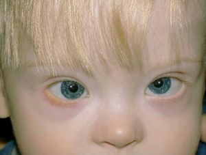
- Eyelid abnormalities including upward slanting palpebral fissures, prominent epicanthal folds, blepharitis, epiblepharon, and congenital ectropion are often seen in Down syndrome (Figure 1).
- The prevalence of slanting palpebral fissures is typically reported at greater than 80%.2
- Epicanthic folds are common and found in greater than 60%.2
- The reported frequency of epiblepharon ranges from 2-54%.2
- The majority of studies reviewed by Creavin et al reported blepharitis in approximately 30%.2
- The increased rate of blepharitis may be related to the narrow, upward slanting palpebral fissures found in these patients in addition to increased susceptibility to infection due to immune system impairment.19
4. Cornea
- Keratoconus (KCN) occurs in up to 1-5% of patients with Down syndrome and typically develops around puberty.10 This may be related in part to steeper than average corneal curvature at baseline compared to age-matched control children.20 Eye rubbing may also contribute to pathogenesis.21 Variants in SOD1 located on chromosome 21, that encodes an superoxide dismutase 1 involved in oxidative stress, have been associated with KCN in 2 families,22,23 although these variants have not been present in other populations with familial KCN.24-26 Whether this represents a genetic basis for a higher prevalence of keratoconus in patients with trisomy 21 is unknown.
5. Lens
- Haugen et al found that the central lens is thinner in patients with Down syndrome (3.27± 0.29 mm) when compared to controls (3.49 ± 0.20 mm) and that the power of the lens in patients with Down syndrome is significantly less (17.70 ± 2.36 D) than that of controls (19.48 ± 1.24 D).8
- Congenital or acquired cataracts occur in approximately 15% of individuals with Down syndrome.27
- The prevalence of cataracts in these patients increases with advancing age. Data has shown that patients with Down syndrome are diagnosed with cataracts at a younger age than individuals in the general population.19
- While early studies showed unfavorable responses to cataract surgery in children with Down syndrome, more recent data has shown no increased rate of surgical complications in these patients when compared to the general pediatric population. In a large case series by Gardiner and colleagues, cataract surgery was performed safely, with good visual outcome. Complications such as aphakic glaucoma were similar to rates of patients without Down syndrome undergoing early cataract extraction.28 However, comorbidities such as congenital heart disease (CHD) must still be considered when placing children under general anesthesia.29,30 If cataracts are deemed amblyogenic in a patient with CHD, a multi-disciplinary pre-, intra- and post-operative plan must be made among the eye surgeon, cardiologist, and anesthesiologist to ensure optimal patient safety. In the senior author’s experience (WWM), cataract surgery for amblyogenic cataracts in the setting of CHD can be performed safely with good outcome, as long as a multi-disciplinary approach is taken.
6. Retina
- Increased retinal vessels crossing the disc margin can be seen in 13-38% of individuals with Down syndrome. 2,31
- Macular hypoplasia has been reported in approximately 1%. 2,5
7. Optic Nerve
- The prevalence of optic nerve elevation has been reported to be 3%. 2
- Optic disc pallor has been reported in 1-5%.2
- Reduced contrast sensitivity has been found in children with Down syndrome when compared to age-matched controls.32
8. Strabismus
- Strabismus occurs in 19-47% of patients with Down syndrome.2,5,8
- The most common type of strabismus is esotropia (84-90%), followed by exotropia (8-10%), and then vertical deviations.8
- Ljubic et al reported the frequency of amblyopia to be 17% in a group of individuals with Down syndrome, which demonstrated the importance of regular eye exams to prevent vision loss from amblyopia.8
9. Visual Cortex
- The layers of the visual cortex have been found to be less organized in patients with Down syndrome when compared to controls.8
- Dendritic spines in the visual cortex have also been shown to be altered and of a lower number than in controls.8
- In addition to the optics of the eye, post-retinal mechanisms have been found to factor into decreased visual acuity in these individuals, using measures of interferometric acuity.8
10. Lacrimal disease
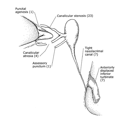
- Nasolacrimal obstruction is found in 17-36% of patient with Down syndrome.12,33 A variety of nasolacrimal duct anomalies have been reported in patients with Down syndrome, including punctal agenesis, canalicular atresia, canalicular and canal stenosis, and anterior placed turbinate (Figure 2).34 A higher rate of failure with simple probing and irrigation surgery is found among this population, and more complex surgical intervention may be required.34,35
- The presence of epiphora has been reported to be 15-32%.2
- Roizen et al reported congenital dacryocutaneous fistulae at a prevalence of 1%.2
11. Nystagmus
- The reported prevalence of nystagmus varies. Creavin et al reviewed 20 studies, 9 of which reported a prevalence between 10-20%, 5 of which reported greater than 20%, and 6 of which reported less than 10%.2
12. Glaucoma
- Glaucoma is found in 1-7% of children with Down syndrome.2
13. Iris
- Brushfield spots in the iris are focal areas of stromal tissue hyperplasia surrounded by relative hypoplasia (Figure 1). They appear as speckled spots and are particularly visible in light irises. They are found in up to 52% of children with Down syndrome and are not of clinical significance.8
Therapeutic Considerations
- Management of medical issues, home environment, education, and vocational training are important considerations that can improve functional prognosis.
- Down syndrome is associated with a shortened life expectancy, with mortality sharply increasing after age 40. However, the life expectancy has significantly increased over the last few decades.
- The leading causes of death are congenital heart disease and infections secondary to impaired immunity, with Alzheimer’s disease being an important secondary cause of death in adults.7
- There is a high rate of early-onset age-related disorders, particularly dementia of the Alzheimer type, musculoskeletal disorders, and sensory impairments. These can cause significant morbidity and should be monitored and treated as they arise.7
- Evaluation for strabismus, cataracts, and nystagmus should begin in the first 6 months of life. Patients should be followed annually until age 5 evaluating for refractive errors, strabismus, or other ocular findings. Dynamic retinoscopy should be performed in patient’s refractive correction to evaluate for hypoaccommodation. Biannual evaluation is then recommended to evaluate for any further ocular manifestations.1
- When treating glaucoma in patients with Down syndrome, medications with cardiovascular or respiratory side effects should be avoided. Likewise, when treating amblyopia, atropine may also have cardiovascular side effects and therefore, methods such as occlusion by patching or optical penalization should be considered.
- Examination technique should take into account cognitive ability, and even adult patients may perform better with recognition acuity testing with LEA symbols or Allen pictures. Teller acuity cards can be utilized when patient is unable to perform recognition acuity techniques. 8

Figure 1. Upward slanting of the eyes, mild esotropia, Brushfield spots on iris, and blepharitis in a young child with Down syndrome.36

Figure 2. Anatomic abnormalities of the nasolacrimal apparatus present in patients with Down Syndrome.34
References
- Bull MJ, Committee on Genetics, Health supervision for children with Down syndrome. Pediatrics. 2011. 128(2):393-406.
- Creavin AL, Brown RD. Ophthalmic assessment of children with Down syndrome: is England doing its bit? Strabismus. 2010. 18(4):142-145.
- Manish N. Approach to Practical Pediatrics. 2011, New Delhi: Jaypee Brothers Medical Publishing.
- Resta RG. Changing demographics of advanced maternal age (AMA) and the impact on the predicted incidence of Down syndrome in the United States: Implications for prenatal screening and genetic counseling. Am J Med Genet A. 2005; 133A(1):31-36.
- Arumugam A. Raja K, Venogopalan M, et al, Down Syndrome-A Narrative Review with a Focus on Anatomical Features. Clin Anat. 2016. 29(5): 568-577.
- Arvind R, Manual of physical diagnosis in pediatrics. 2016, New Delhi: Jaypee Brothers Medical Publishers.
- Torr J, Strydom A, Patti P, Jokinen N. Aging in Down Syndrome: Morbidity and Mortality. Journal of policy and practice in intellectual disabilities. 7(1):70-81.
- Watt T, Robertson K, Jacobs RJ. Refractive error, binocular vision and accommodation of children with Down syndrome. Clin Exp Optom. 2015. 98(1):3-11.
- Doyle L, Saunders KJ, Little JA, Determining the relative contribution of retinal disparity and blur cues to ocular accommodation in Down syndrome. Sci Rep. 2017. 7:39860.
- Paudel N, Leat SJ, Adhikari P, Woodhouse JM, Shrestha JB, Visual defects in Nepalese children with Down syndrome. Clin Exp Optom. 2010; 93(2):83-90.
- Fong AH, Shum J, Ng AL, Li KK, McGhee S, Wong D. Prevalence of ocular abnormalities in adults with Down syndrome in Hong Kong. Br J Ophthalmol. 2013; 97(4):423-428.
- Haugen OH, Høvding G, Lundström I, Refractive development in children with Down's syndrome: a population based, longitudinal study. Br J Ophthalmol. 2001; 85(6):714-719.
- Garcia ML, Huang D, Crowe S, Traboulsi EI. Relationship between the axis and degree of high astigmatism and obliquity of palpebral fissure. J AAPOS. 2003; 7(1):14-22.
- Knowlton R, Marsack JD, Leach NE, Herring RJ, Anderson HA. Comparison of Whole Eye versus First-Surface Astigmatism in Down Syndrome. Optom Vis Sci. 2015. 92(7):804-814.
- Marsack JD, Ravikumar A, Benoit JS, Anderson HA. Variability in Objective Refraction for Persons with Down Syndrome. Optom Vis Sci. 2017. 94(5):574-581.
- Hunter DG. Dynamic retinoscopy: The missing data. Surv Ophthalmology. 2001; 46(3):269-274.
- Cregg M, Woodhouse JM, Pakeman VH, et al. Accommodation and refractive error in children with Down syndrome: cross-sectional and longitudinal studies. Invest Ophthalmol Vis Sci. 2001; 42(1):55-63.
- Nandakumar K, Evans MA, Briand K, Leat SJ. Bifocals in Down syndrome study (BiDS): analysis of video recorded sessions of literacy and visual perceptual skills. Clin Exp Optom. 2011; 94(6):575-585.
- Krinsky-McHale SJ, Jenkins EC, Zigman WB, Silverman W. Ophthalmic disorders in adults with down syndrome. Curr Gerontol Geriatr Res. 2012. 2012:974253.
- Vincent AL, Weiser BA, Cupryn M, Stein RM, Abdolell M, Levin AV. Computerized corneal topography in a paediatric population with Down syndrome. Clin Exp Ophthalmol. 2005; 33(1):47-52.
- McMonnies CW. Mechanisms of rubbing-related corneal trauma in keratoconus. Cornea. 2009; 28(6):607-615.
- Udar N, Atilano SR, Small K, Nesburn AB, Kenney MC, SOD1 haplotypes in familial keratoconus. Cornea. 2009; 28(8):902-907.
- Udar N, Atilano SR, Brown DJ, et al SOD1: a candidate gene for keratoconus. Invest Ophthalmol Vis Sci. 2006; 47(8): 3345-3351.
- Nejabat M, Naghash P, Dastsooz H, Mohammadi S, Alipour M, Fardaei M. VSX1 and SOD1 Mutation Screening in Patients with Keratoconus in the South of Iran. J Ophthalmic Vis Res. 2017; 12(2): 135-140.
- Al-Muammar AM, Kalantan H, Azad TA, Sultan T, Abu-Amero KK. Analysis of the SOD1 Gene in Keratoconus Patients from Saudi Arabia. Ophthalmic Genet. 2015; 36(4):373-5.
- Stabuc-Silih M, Strazisar M, Hawlina M, Glavac D. Absence of pathogenic mutations in VSX1 and SOD1 genes in patients with keratoconus. Cornea. 2010; 29(2):172-176.
- Cuniff C, Trotter T. Health supervision for children with Down syndrome - Reply. Pediatrics. 2001; 108(6):1385-1385.
- Gardiner C, Lanigan B, O'Keefe M. Postcataract surgery outcome in a series of infants and children with Down syndrome. Br J Ophthalmol. 2008; 92(8):1112-1116.
- Saifee M, Kong L, Yen KG. Outcomes of Cataract Surgery in Children with Down Syndrome. J Ophthalmic Vis Res. 2017; 12(2):243-244.
- Li EY, Chan TC, Lam NM, Jhanji V. Cataract surgery outcomes in adult patients with Down's syndrome. Br J Ophthalmol. 2014; 98(9):1273-1276.
- Kim JH, Hwang JM, Kim HJ, Yu YS. Characteristic ocular findings in Asian children with Down syndrome. Eye (Lond). 2002; 16(6):710-714.
- John FM,Bromham NR, Woodhouse JM, Candy TR. Spatial vision deficits in infants and children with Down syndrome. Invest Ophthalmol Vis Sci. 2004; 45(5):1566-1572.
- Berk AT, Saatci AO, Erçal MD, Tunç M, Ergin M. Ocular findings in 55 patients with Down's syndrome. Ophthalmic Genet. 1996; 17(1):15-19.
- Coats DK, McCreery KM, Plager DA, Bohra L, Kim DS, Paysse EA. Nasolacrimal outflow drainage anomalies in Down's syndrome. Ophthalmology. 2003;. 110(7):1437-1441.
- Lueder GT. Treatment of nasolacrimal duct obstruction in children with trisomy 21. J AAPOS. 2000; 4(4):230-232.
- Traboulsi EI, A compendium of inherited disorders and the eye. Ophthalmology monographs. 2006, New York: Oxford University Press in cooperation with the American Academy of Ophthalmology.