Researchers at Vanderbilt University have discovered the mechanism that allows zebrafish to regenerate their retinas and recover vision after injury.
The NEI-funded study published in Stem Cell Reports, found that when retinal cells die they suppress signaling of the neurotransmitter gamma-aminobutyric acid (GABA), which in turn induces Müller glia to return to a stem cell-like state and proliferate throughout the area of injury.
Previous investigations have shown that GABA activity in the hippocampus and mouse pancreas modulates stem cell activity. When GABA levels are high, the stem cells remain largely inactive, but when levels decline, the cells begin to divide.
Based on these findings, senior author James Patton, PhD, and his student Mahesh Rao injected GABA inhibitors into the undamaged retinas of zebrafish. They found that GABA suppression caused spontaneous proliferation of Müller glia-derived progenitor cells, whereas increasing GABA signaling after an induced retinal injury inhibited the regenerative response by reducing proliferation.
Biologists of different disciplines have been studying zebrafish for years due to their unique regenerative abilities. In addition to their retinas, larval zebrafish can regrow and repair their fins, skin, heart, lateral line sensory organ and brain. The diminutive tropical fish, well-known to many as a cheerful and hardy aquarium species, were one of the first vertebrates to be cloned, and are used as model organisms in a variety of research.
The authors hope these findings may soon be applied to create alternative therapies for degenerative retinal diseases such as retinitis pigmentosa. Currently, there are several treatment candidates in development that use injections of stem cells or retinal precursors. However, none have shown efficient integration into existing neural circuits or success at restoring vision in humans.
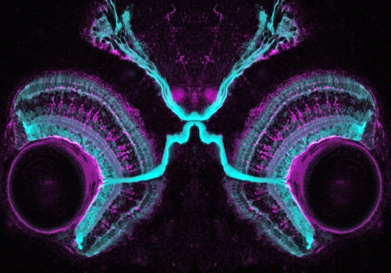
 Image courtesy of Dr Kara Cerveny & Dr Steve Wilson, Wellcome Images | Confocal micrograph showing the connections of the visual system in a four-day-old zebrafish embryo. Both the retinal ganglion cell layers and photoreceptor cell layers are shown in cyan, the glial cells are shown in purple.